background image


INTRODUCTION


Thank you for your purchase of a National microscope.  It is a well built, precision instrument carefully checked to 
assure that it reaches you in good condition.  It is designed for ease of operation and years of carefree use.  The 
information in this manual probably far exceeds what you will need to know in order to operate and maintain your 
microscope.   However, is provided to answer questions which might arise, and to help you avoid any maintenance 
expense that may be unnecessary. 

 
Your new compound microscope is a high performance microscope with high quality achromatic objective lenses 
that provide good resolution and optical centering.  The microscope is designed with a built-in ball bearing 
mechanical stage providing a travel range of 75mm x 50mm in the X and Y direction with graduation reading up to 
0.1mm for accurate positioning of specimen.  Also included is a ball bearing quadruple nosepiece, precision coaxial 
focusing mechanism, rack and pinion mounted N.A. 1.25 Abbe condenser and built-in 3 watt LED variable light 
source.  

Retain the styrofoam container in case the microscope must be transported or returned to factory 

for any reason. 

 
Carefully read these instructions before operating microscope.  They will permit you to use your new microscope to 
its fullest capability.  Nomenclature used to describe components and controls is identified by referring to diagram 
on page 2.


UNPACKING


The microscope and accessories have been carefully packed to assure they reach you in the best possible 
condition.  Do not discard the packing container or materials until all components are accounted for.  Save the 
packing container in case the microscope needs transporting to another location or shipped for repairs.  
Components are packed within the container as indicated below. 


167 Monocular: 

Head and Stand, one eyepiece, one rubber eyeshield, 12VDC switching power supply, 
operates on 100v-240v, 50H/60H, and dust cover. 

168 Binocular head 

Head and Stand, two eyepieces, two rubber eyeshields, 12VDC switching power 
supply, operates on 100v-240v, 50H/60H, and dust cover. 

169 Trinocular head 

Head and Stand, two rubber eyeshields, c-mount, 12VDC switching power supply, 
operates on 100v-240v, 50H/60H, and dust cover. 


A. 

Lay container (A) flat and carefully remove microscope head and stand. 


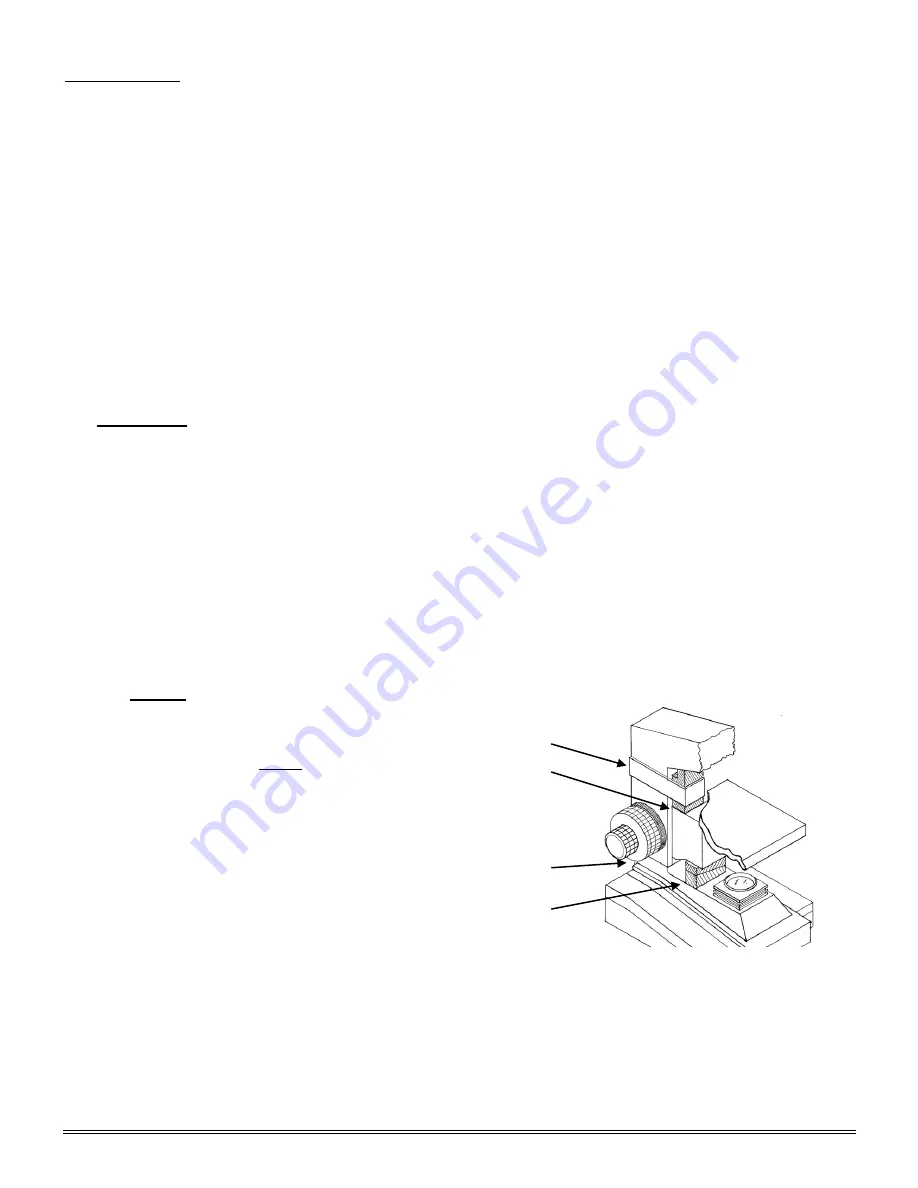
NOTICE

 To protect focus mechanism during 


shipment, two black plastic wedges (b)  


and one black plastic block (c) are 


inserted at strategic points as indicated. 
These plastic parts 

MUST

 be removed prior 

to operating microscope.  Failure to do so 
will result in damage to focusing mechanism 
and will void your warranty. 
 
1.  Remove two black Velcro straps (a). 
2.  Remove wedges (b) by pulling apart the 

            two parts of wedge in opposite directions.    

               3.   Lower stage by rotating coarse focus 


             knob, on side of microscope illustrated,        
             in counter-clockwise direction 
        4.  Remove blocks (c) from stand. 
        5.  These components should be retained with styrofoam container 


B. 

Carefully remove from the stand all tape and packing material used to protect microscope components during 
shipment.  
 

C.  Un-wrap the components, making certain that lens surfaces do not come in contact with dust, dirt, 

fingerprints.   Damage to optical surfaces can result from such contaminants, and reduce image quality. 


(a) 

(b) 

(a) 

(c) 

Summary of Contents for 167 Series

Page 1: ...ts Inc 6508 Tri County Parkway Schertz Texas 78154 Phone 210 590 9010 Fax 210 590 1104 INSTRUCTIONS FOR 167 168 169 DC20 169 and 169 BTW1 Series COMPOUND BIOLOGICAL MICROSCOPES Copyright 11 4 2016 National Optical Scientific Instrument Inc ...

Page 2: ...ges Software MAC Full Help Menu 16 Introduction to the 169 BTW1 18 169 BTW1 Assembly Connecting Camera to Tablet 19 Tablet Operation Power Management 20 Tablet Accessories 21 Camera Switches and Ports Connecting to WiFi device Connecting to WiFi Computer 22 Connecting to Existing WiFi Network 23 Connecting to Networked BTW with MotiConnect App 25 Connecting to Networked BTW with Motic Images 27 Ta...

Page 3: ... condition Do not discard the packing container or materials until all components are accounted for Save the packing container in case the microscope needs transporting to another location or shipped for repairs Components are packed within the container as indicated below 167 Monocular Head and Stand one eyepiece one rubber eyeshield 12VDC switching power supply operates on 100v 240v 50H 60H and ...

Page 4: ...rtical port Retighten knurled screw to secure vertical eyepiece tube in place 2 Trinocular head Mounting C Mount to head Loosen knurled locking screw remove black dust cap from vertical port Insert C Mount and retighten knurled screw to secure in place a Trinocular head provided with a three position sliding rod to direct light through microscope The three position sliding rod b allows user to eas...

Page 5: ...ry adjustment of viewing head Models 168 and 169 only 1 Look through microscope and adjust distance between the two eyepiece tubes by grasping the sliding mounts to left and right of eyepieces and sliding together or apart 2 When a full field of view is observed through both tubes and images blend into one interpupillary distance is corrected for your eyes Check the interpupillary scale and note i...

Page 6: ...care must be exercised in order to prevent damaging the front lens element and specimen slide d In order to obtain maximum resolution of the 100x oil immersion lens it is necessary to apply immersion oil between the coverglass of slide and front lens of the objective 1 Use of a very small amount of immersion oil is required Only the very tip of the lens should ever come in contact with the immersi...

Page 7: ...he top CS adaptor ring in order for the video adaptor to be compatible with the chip size of your digital c mount camera MAINTENANCE WARNING FOR YOUR SAFETY TURN SWITCH OFF AND REMOVE PLUG FROM POWER SOURCE OUTLET BEFORE MAINTAINING YOUR MICROSCOPE TO AVOID SHOCK OR FIRE HAZARD IF POWER CORD IS WORN CUT OR DAMAGED IN ANY WAY HAVE IT REPLACED AT ONCE A OPTICAL MAINTENANCE 1 Do not attempt to disass...

Page 8: ...r located on top of mechanical stage b Remove 5ea cross head screws holding base plate c Carefully lay the base plate flat on the table d Disconnect the LED cable from the plug socket on the LED circuit card e Your microscope requires a 3W LED bulb assembly Part 800 500 f The LED bulb is located within the lamp housing It is held in place with screw on holder Simply unscrew the ring holder to remo...

Page 9: ...1080 Live Display Mode through USB 1980 x 1080 Live Display Mode through HDMI 1920 x 1080 HD 60 fps Pixel Size 2 8μm x 2 8μm Data Transfer HDMI 1080P and USB2 0 Operating Temperature From 10 to 60 Degrees Celsius non condensing Slot SD Card max 32 GB Supported OS Microsoft Windows XP Vista 7 8 and MAC OSX Minimum Computer Requirements 2GHz dualcore RAM mem 2GB Video mem min 512 MB ...

Page 10: ...10 DC20 169 Series ASSEMBLY ...

Page 11: ...be used to connect a USB mouse so that the camera can be controlled without a computer The USB connection can be used to connect the Moticam 1080 directly to a computer not recommended to be used with the provided Motic Images Plus software HDMI port for connecting to an HDMI ready display the cable is provided in the Moticam 1080 box DC 12V Power port for connecting the included Power Supply SD C...

Page 12: ...d now illuminate 4 Now make sure your microscope is on and you are focused in on your slide specimen 5 On the right hand side of the head just behind the right eyepiece is the three position slide bar The slide bar must be pulled out until it stops 6 In this position the slide should be visible through the eyepieces and displayed on your HD ready device ...

Page 13: ...splay for capturing images and also for adjusting various aspects All images can be captured to 32GB Max SD card for transfer to computer later Live Image and Capture Controls 1 Zoom In Zooms in on an area of the live image 2 Zoom Out Zooms out on an area of the live image 3 Magnifier Zooms in full screen a selected region of live image 4 Region of Interest Allows user to view only a specific area...

Page 14: ...posure Gain Gamma Brightness Light Frequency White Balance and Color Temp Settings Menu allows you to adjust Saturation Contrast Gamma Sharpness Digital Noise Save Individual User settings Time setting Language Format the SD Card and Restore all settings to Default ...

Page 15: ...er images to your computer Images are automatically captured as Jpeg images Please note 32GB Max MOTIC IMAGES SOFTWARE PC Full Help Menu The full software manual for Motic Images is accessible within the software s main page To begin open the Motic Images Software At the top of main screen find the menu tab labeled Help Click on Help and then select the help option This will open the Motic Images ...

Page 16: ...eled File and click on Capture Once the Motic Live Imaging Module has opened click on Help This will open the Motic Live Imaging Module help file containing the full help menu MOTIC IMAGES SOFTWARE MAC Full Help Menu The full software manual for Motic Images is accessible within the software s main page To begin open the Motic Images Software At the top of main screen find the menu tab labeled Mot...

Page 17: ...17 This will open the Motic Images Help Manual ...

Page 18: ...or 5 0 MP captured image resolution WiFi Resolution 2 0MP 5 0MP 720P captured video resolution HDMI 1080 Android operating system WiFi WiFi output 802 1 ab g n supporting both hot spot mode or using existing WiFi network Preloaded optimized MotiConnect app Supplied with Motic Images Plus software for Mac PC Built in MoticHub allows for wireless sharing ...

Page 19: ...al USB cable cord and plug one end into your camera and the other into the side of the Tablet screen See picture above 3 Unless your tablet is fully charged plug the power adapter into the back of the camera next to the USB port 4 This will both charge and allow you to use the camera tablet at the same time 5 Unplugged from the charger the tablet camera are able to function for about 2hrs ...

Page 20: ...wo hours of continuous use is required use the supplied wall charger and power cable included with your tablet Leaving the power charger connected to the camera will trickle charge the tablet This will be a much slower rate but should allow time beyond 2hrs 2 Camera connected to both tablet and wall charger Trickle charging tablet via USB Volume adjustment decrease Volume adjustment increase Hold ...

Page 21: ...A15602 C Mount 4GB Micro SD This storage card can be used with your tablet to store images and then transfer them to your computer This card is provided as a courtesy and cannot be replaced 32GB Max Supported Mini USB to Micro USB Provides power and feed connection between the tablet and camera To operate the camera move the switch to the camera position USB to Micro USB This cable when used in co...

Page 22: ... camera to the power supply 3 Switch the camera to WiFi transmission mode 4 Turn on your WiFi enabled device and connect to the SSID provided on the camera MCX_BTW_XXXX You will connect to the camera as you would any WiFi router 5 Default password is always 12345678 6 Start the MotiConnect application The connection should be automatic 7 You can now view the image live on the MotiConnect app as we...

Page 23: ... existing network Advanced The BTW camera has the unique ability to connect to your existing network system using the MoticHub feature 1 On the home screen of the tablet select the Settings icon 2 Select WiFi within the Settings window ...

Page 24: ...ect turn WiFi on if turned off Note In this case I have selected National 4 Now you have added the tablet to your WiFi network 5 On the home screen of the tablet select the MotiConnect app 6 Then select the MoticHub icon and MoticHub will launch ...

Page 25: ...onnect your WiFi enabled device tablet phone computer laptop 10 Now any tablet or computer laptop connected to your network will have access to your live image Connecting to Networked BTW with MotiConnect App 1 Make sure you are first connected to your office home WiFi network 2 Download the MotiConnect app from the App Store Google Play or Chrome Store to your android or apple device tablet phone...

Page 26: ...ice button at the top right hand of the screen 5 Click on the Add button on the pop up window 6 You may give your New Device any name however the IP Address must be same as the IP generated by MoticHub 7 Now select your New Camera ...

Page 27: ...ture measure and annotate the image using the tools provided Connecting to Networked BTW with Motic Images 1 Install the Motic Images software into your computer or laptop 2 Start the Motic Images application Click File Click Capture 3 The Motic Live Imaging window will open ...

Page 28: ... X selected 5 Click on Open to open the Moticam X IP address box 6 In the open Moticam X IP address box type in the IP address generated by the MoticHub feature of the BTW tablet 7 Once you click OK the image produced by your BTW tablet should appear ...

Page 29: ...he primary application for accessing your camera is MotiConnect T MOTICONNECT T OVERVIEW HELP MENU MotiConnect is a dedicated image processing Android app for Motic cameras which includes image preview capture recording editing and measuring functions Please refer to the Application Help guide for a complete description of the MotiConnect Application HELP BUTTON Help Menu Opens a new window which ...

Page 30: ...fication window select the objective you captured the dot with In this case the 4X objective 7 In the diameter window select the diameter of the dot you captured In this case the 150um Then click OK 8 The last window that will open is the Save Calibration window Since you capture this image with the 4X objective it is best to enter this on this line 9 The objective line should default to the objec...

Page 31: ... file your images Full Help Menu The full software manual for Motic Images is accessible within the software s main page To begin open the Motic Images Software At the top of main screen find the menu tab labeled Help Click on Help and then select the help option This will open the Motic Images help file contents containing the full help menu ...

Page 32: ...pture your images Full Help Menu The full Live Imaging Module manual is accessible within the live Imaging main page To begin open the Motic Images Software At the top of main screen find the menu tab labeled File and click on Capture Once the Motic Live Imaging Module has opened click on Help This will open the Motic Live Imaging Module help file containing the full help menu ...

Page 33: ...e are help files for both the main Motic Images software window as well as the Motic Images Live Imaging window MOTIC IMAGES 3 0 HELP GUIDE To access the Motic Images 3 0 help menu click on Help located at the top of the Motic Images software screen Once the Help window open you will find the help guide within Motic Live Imaging Module Help To access the Motic Images Live Imaging help menu click o...

Page 34: ...nser lens a Blue filter is utilized for bright field observation b Green filter is utilized for phase observation C Install objectives 1 Rotate coarse focusing knob to move microscope stage platform to its highest position 2 Remove objective dust caps from the revolving nosepiece 3 Screw objective lenses into nosepiece making certain to mount them in consecutive order 10x 20x 40x and 100x OPERATIO...

Page 35: ...ulus ring annuli located in phase turret condenser until it is centered to the larger dark annulus located in the objective Both rings must be concentric to each other to achieve maximum performance Make sure that the knurled head centering screws are disengaged from the hex socket screws of annuli centering mechanism and in the out position before rotating phase turret condenser R Repeat above st...

Page 36: ...pe such as mirrors lamps filters intermediate lenses In the case of microscopes with a camera attached to it Dirt on the camera adapter Dirt on the protection filter of the camera sensor For Eyepieces with reticules Dirt on the outer or inner reticle glass Objectives are the optical component of the microscope that require the most maintenance Because for their actual use they can get dirty easily...

Page 37: ...ponents of the microscope such as the ON OFF switch the dimmer the lamp holder If there are grease stains use the same cloth moistened with a low graduation of alcohol If you face any problems related to the maintenance of your microscope please contact us Our technicians will gladly help you solve your maintenance issue s CLEANING The front lens of the objectives particularly the 100XRD should be...

Page 38: ... holder Have qualified service technician repair outlet IMAGE QUALITY PROBLEM REASON FOR PROBLEM SOLUTION No image Nosepiece not indexed properly Light too bright Move revolving nosepiece until objective lens clicks into position Adjust light intensity control to a lower position Poor resolution Image not sharp Objective lens dirty Eyepiece lens dirty Slide upside down Cover slip on specimen slide...

Page 39: ...IN 40X Plan objective lens 0 65 N A 760 160P DIN 60X Plan objective lens 0 90 N A 799 160P DIN 100X Plan objective lens 1 25 N A 800 500 BULB ASSEMBLY 3W LED 926 Phase contrast set Centering telescope PLAN Phase 10x 20x 40x 100xR oil immersion objectives Phase turret condenser assembly including one brightfield position blue green filters storage case 965 160 Eyepiece reticle 10mm 100 div 975 002 ...

Reviews: