57
4.11 Examinations in incident light brightfield,
darkfield, polarisation contrast, interfer-
ence contrast
Please proceed as follows for selecting and set-
ting contrasting techniques:
Incident light brightfield:
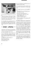
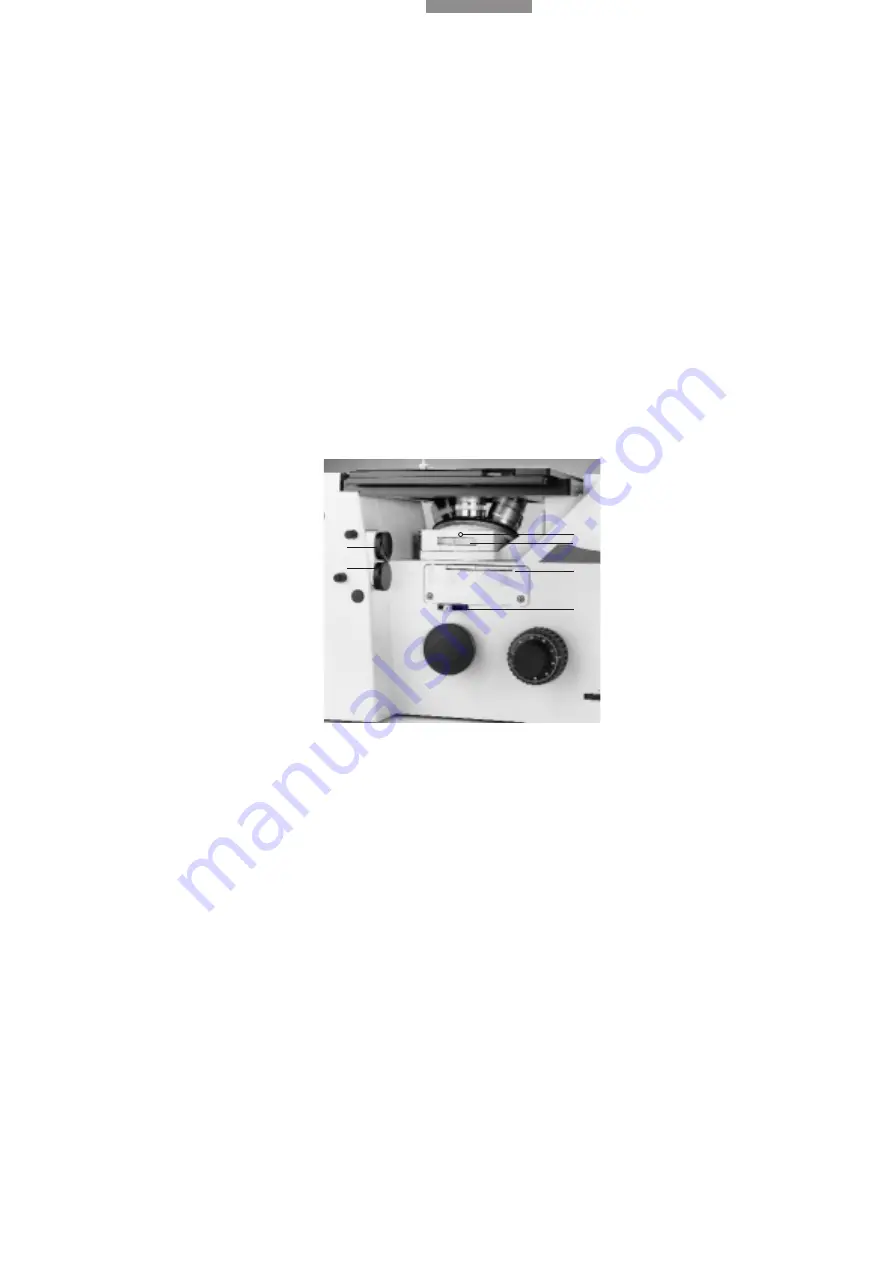
Turn a BF or Smith reflector into the light path by
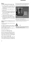
rotating the turret (76.1).
If using gas discharge lamps or if looking at
strongly reflecting surfaces or switching quickly
between brightfield and darkfield, it is advisable
to slot an N16 neutral density filter (29.2) into the
ring mount of the BF reflector (but not recom-
mended for polarisation contrast and interfer-
ence contrast).
Rotate the ICR prism turret (if used) to the BF
position to disengage the ICR prisms.
Pull or swing the polariser, analyser, ICR prism
out of the light path.
Turn a low magnification into the light path and
adjust the illumination to a medium setting.
Make sure the front lens of the objective is clean,
close the field diaphragm (76.4) half way, open
the aperture diaphragm (76.5).
Focus the specimen surface, the half-closed field
diaphragm makes it easier to find the specimen
surface.
Open the field diaphragm until it just disappears
beyond the edge of the field of view. The setting
of the field diaphragm remains the same for all
objective magnifications.
The aperture diaphragm influences the resolution,
contrast and field depth of the microscope image.
It should be opened just wide enough to encom-
pass the entrance pupil of the objective (brighter
circle) and to cut off stray light at the walls of the
microscope.
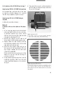
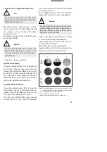
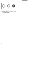
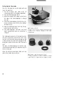
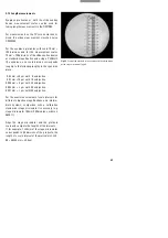
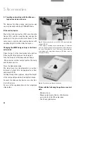
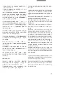
Then the illumination aperture is equal to the
objective aperture (77.1).
This can be checked by looking through the
empty eyepiece tube after removing one of the
eyepieces (77). If included in the configuration,
the Bertrand lens can be engaged for magnified
viewing.
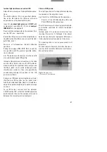
In general the aperture diaphragm is varied
according to the visual impression of the speci-
men, contrast and field depth. We recommend
closing the aperture diaphragm by 1/3 (77.2).
Narrowing the aperture diaphragm too far results
in pronounced diffraction phenomena at weak
and medium objective magnifications.
With high aperture, high-magnification objec-
tives the aperture diaphragm can be narrowed
further, which generally leads to a major im-
provement in contrast (77.3).
The aperture diaphragm must not be used for
adjusting the image brightness.
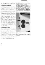
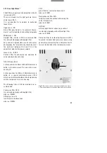
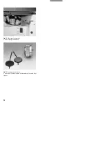
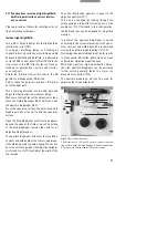
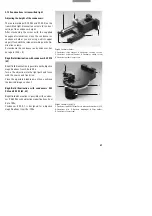
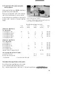
Fig. 76
Side view of microscope
1
Reflector turret,
2
ICR turret,
3
Analyser (polariser on other
side of microscope),
4
Field diaphragm,
5
Aperture diaphragm,
6
Position of the knurled knob for ICR contrast setting
1
3
5
4
2
6


