Levenhuk 670T: Microscope body, trinocular head, achromatic objective lenses (4x, 10x, 40x S,
100x S (oil immersion)), WF10x and WF20x eyepieces (2 pcs), 6V/20W halogen lamp, blue filter,
vial of immersion oil, dust cover.
Levenhuk D670T: Microscope body, trinocular head, achromatic objective lenses (4x, 10x, 40x S,
100x S (oil immersion)), WF10x and WF20x eyepieces (2 pcs), 6V/20W halogen lamp, blue filter,
vial of immersion oil, 5 mpx digital camera, software & drivers CD, dust cover.
Levenhuk 625 / Levenhuk 670T / Levenhuk D670T is comprised of nine parts:
Base: not only does it hold the weight of the microscope, it also houses the illumination source,
electronics and control mechanisms.
Arm: this piece holds the base, the stage and the head of the microscope together. Coarse and
fine focus systems provide for smooth vertical movements of the stage.
Rack-and-pinion mechanism: mounted on the arm, the stage with the condenser are moving
vertically along this column. For additional precision, a condenser may be adjusted separately.
Head: a binocular (Levenhuk 625) or a trinocular (Levenhuk 670T / Levenhuk D670T) head is
mounted at a 30° angle at the upper end of the arm.
Eyepieces: wide FOV eyepieces WF10x and WF20x are used in these microscopes.
Revolving nosepiece: quadruple revolving nosepiece allows you to change objective lenses
smoothly and easily.
Objective lenses: high-quality achromatic objective lenses with 4x, 10x, 40x and 100x
magnifications provide for sharp and bright images.
Stage: sturdy and reliable stage with a specimen holder that can be used to move your slides
while observing them.
Condenser: Abbe condenser, with 1.25 N.A. iris diaphragm.
Parts of the microscope
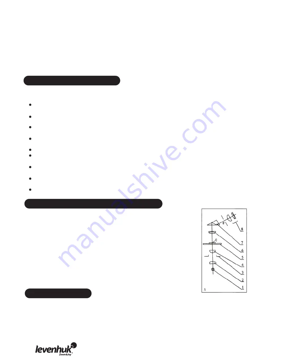
Operating principle and illumination
1. Image creation system: objective lens (6), prism (7) and eyepiece (9).
The objective lens (6) magnifies a specimen (0), light rays pass through a
prism (7), refract at a 45
о
angle and create an image in the eyepiece.
Total magnification may be calculated by multiplying magnifications of
the eyepiece and the objective lens used.
2. Illumination system: lamp (1), collector lens (2), diaphragm (3) and
condenser (4). Light emitted from a lamp (1) passes through a collector
lens (2) and illuminates a diaphragm (3). After this, it is focused by a
condenser (4). This illumination system is used for observations of a
specimen (0) in transmitted light. However, you can also use a different
type of illumination for observations in reflected light.
Levenhuk D670T comes with a 5 mpx C510T NG digital camera.
The camera allows you to observe specimens in fine detail and true colors on your PC monitor
and save images on the hard drive.
The special software that comes in the kit allows you to view and edit the resulting images.
Supported file formats include: *.bmp, *.jpg,*.jpeg,*.png, *.tif, *.tiff, *.gif, *.psd, *.ico, *.emf,
*.wmf, etc.
Digital camera


