background image


ZS3 Ultrasound System


Instructions for Use


Summary of Contents for Zonare ZS3

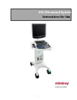
Page 1: ...I ZS3 Ultrasound System Instructions for Use ...

Page 2: ...9 781 8 002 705 8 226 561 D461 814 D462 446 D467 002 D469 539 D469 877 SHENZHEN MINDRAY BIO MEDICAL ELECTRONICS CO LTD Mindray Building Keji 12th Road South High Tech Industrial Park Nanshan Shenzhen 518057 P R China Shanghai international Holding Corp GmbH Europe Eiffestraβe 80 20537 Hamburg Germany Technical Support North America and Asia Phone support 1 877 913 9663 or 1 650 316 3199 option 7 u...

Page 3: ...ers 66 7 Archiving Review and Management 69 8 AdvancedFeatures and Options 76 Contrast Enhanced Ultrasound CEUS 76 Contrast Enhanced Ultrasound Quantification Imaging CEUS QI 80 Elastography 83 Tissue Tracking Quantitative Analysis TTQA 89 9 3D 4D Imaging with Advanced Lighting 92 10 Needle Optimization Mode For Improved Needle Visualization 99 11 Protocols 101 12 Stress Echo 103 13 Setup 107 14 S...

Page 4: ...he property of their respective owners This posting serves as notice under 35 U S C 287 a for Mindray patents http www mindrayna com patents Responsibility on the Manufacturer Party Contents of this manual are subject to change without prior notice All information contained in this manual is believed to be correct Mindray shall not be liable for errors contained herein or for incidental or consequ...

Page 5: ...of the product or the use of parts or accessories not approved by Mindray or repairs by people other than Mindray authorized personnel This warranty shall not extend to Malfunction or damage caused by improper use or man made failure Malfunction or damage caused by unstable or out of range power input Malfunction or damage caused by force majeure such as fire and earthquake Malfunction or damage c...

Page 6: ......

Page 7: ...ilability of application may be limited based on country or region of use Device Description The ZS3 Ultrasound Platform and products derived there from for example but not limited to the ZS3 and the z onepro with and without the SP UI option is used for ultrasound evaluation of the following applications Laparoscopic Fetal Abdominal Intra operative Pediatric Ophthalmic Small Organ Parts breast te...

Page 8: ...nly applicable for liver in the FDA region 9 Cardiac CEUS Only applicable for LVO in the FDA region 10 CEUS QI Only applicable for liver and LVO in the FDA region 11 CEUS Sequence Only applicable for liver and LVO in the FDA region 12 HD Scope 13 Advanced Cardiac Including AutoEF and Expanded Echo Calcs 14 Smart OB NT 15 AutoIMT 16 Stress Echo 17 Total Recall 18 Protocol 19 Needle OPT 20 Anat Mmod...

Page 9: ...pported Item Model Digital black white video printer SONY UP D711MD Digital color video printer SONY UP D25MD Note The availability of options may be limited based on country or region of use The following symbols may be used in this document or elsewhere in product labeling Symbol Description Information that may relate to safety of the patient the operator or the equipment A type BF patient appl...

Page 10: ...re information on returning or recycling this system please contact Shenzhen Mindray Bio Medical Electronics Co or the distributor from whom you purchased the system Consult the Instructions for Use Serial number Authorized representative in the European Community Catalog number Shipping Storage Fragile Shipping Storage Keep dry Shipping Storage Temperature limits Shipping Storage This side UP Shi...

Page 11: ...rsion for 30 minutes at a depth of 1 meter This product is provided with a CE marking in accordance with the regulations stated in Council Directive 93 42 EEC concerning Medical Devices The number adjacent to the CE marking 0123 is the number of the EU notified body certified for meeting the requirements of the Directive MR Unsafe the system is not intended to be used within magnetic resonance MR ...

Page 12: ......

Page 13: ...erview of Console System and Features 19 inch display Gel Bottle Holder Storage Bins Cable management hooks 2 USB Ports Aux CW Port Speakers User Interface Control Panel height adjust Power button Multi Transducer Port MTP ECG Connector Three wheels have a brake function One Front swivel lock Wheel Figure 2 1 ZS3 System Components ...

Page 14: ...ately by using the Pointer to click on the Full Image Display ICON located in the lower left corner of the imaging screen Once in Full Image Display the screen will display Thumbnails or F Keys OLEDs and Small Thumbnails Use the Pointer to click on the Switch Button ICON on the far right hand side of the screen to toggle between F Keys and OLEDs Click on Thumbnail ICON to toggle between large and ...

Page 15: ... is 128 GB NOTE The internal hard drive is not intended for use as a long term archive Back up of exam data is recommended regularly USB Ports Four USB ports two in the front and two in the back allow importing exporting of exams to from the system as well as to install software and to backup restore system and preset preferences Note Before deleting any exam data from the cart hard drive always v...

Page 16: ... hours of normal use in the absence of a connection to an active AC power outlet depending on usage The battery allows the system to be operated in normal use without connection to an active AC Power outlet The backup is mounted at the base of the system and is automatically kept charged as needed whenever the ZS3 system is connected to active AC power Printers and Other Peripherals Mindray offers...

Page 17: ...wrapped over the cable hooks 4 Place wheel s in the non swivel position 5 Ensure that all wheels do not have the brakes set ON and push the cart from the front when transferring from location to location 6 When moving the cart over obstacles always pull from the front Before Scanning 1 Before scanning a patient apply the brake by fully depressing any of the three brake pedals 2 To disengage brakes...

Page 18: ...ZS3 Instructions for Use 11 P a g User Interface and Onscreen Display Figure 2 4 Keyboard Figure 2 5 User Interface See Section 3 Imaging for more details ...

Page 19: ...ZS3 Instructions for Use 12 P a g e Patient and Institution Information Menus Image Area Image Parameters Trackball Priority ICON OLEDs Programmable Keys ...

Page 20: ...on and protocol information Trackball and set keys are used to navigate this area Image Area Displays the ultrasound images Doppler spectral strip M Mode strip image depth markers ECG and respirometer waveforms probe indicator and calculations measurement results window Image Parameters Displays the image parameter for the active window If there is more than one imaging mode active the parameters ...

Page 21: ...ifferent subset of controls Thumbnails Thumbnail Navigation Arrows and Thumbnail Sizing Displays thumbnails of the images stored on the current patient Images can be viewed edited deleted or sent to media by pressing the pointer key and then navigating the pointer to the desired thumbnail and clicking on them Large arrows on either end of the thumbnail strip can be clicked to navigate through the ...

Page 22: ...n 1 Press Caliper while image is frozen 2 Select desired measurement tool on menu press a Set Key 3 Position first caliper with Trackball press a Set Key Position second caliper with Trackball press a Set Key Note Pressing the Select Key will toggle between active calipers in the set or series for fine tuning Basic Calculations Calc Button 1 Press Calc while image is frozen 2 Select desired calc o...

Page 23: ...down menu This will launch a page to select the destination 4 Select DICOM Media from the list 5 Select the Destination 6 Select OK at bottom of screen Review Images in Current Exam 1 Press Current Exam back row of QWERTY keyboard 2 All stored images for exam appear in a Multi Image Format 3 To view an image in Full Image with Thumbnails format point with the cursor and Double Click a Set key 4 To...

Page 24: ...the thumbnail to be reviewed 3 To delete the image point to the trashcan graphic and press Set A prompt will ask to confirm Click OK 4 To view the image in the image area Double Click Set key 5 To return to live imaging press Exit Review OLED or Current Exam again ...

Page 25: ...d exits to live scanning Cancel button exits page Basic Operating Controls Control Description Use Transducer Button Press the Transducer button to select one of three imaging or one non imaging probes attached to the system using the softkey OLEDs Exam Type Button Press the Exam Type button to select create modify and delete presets 11 presets per probe can be assigned to sofkey OLEDs for easy ac...

Page 26: ...tween active calipers in a set to fine tune while Set will continue to launch calipers and complete enter calculations When in color and spectral Doppler Select will toggle through sizing and positioning the color ROI as well as moving the PW cursor while the image is live while Set will continue to toggle between the 2D image and the spectral Doppler When Image Width and Color Doppler are both ac...

Page 27: ...user can press the Priority button to toggle to Color Doppler if turned ON for instance which will change the OLEDs and Onscreen Trackball Priority ICON to reflect Color Doppler controls without having to turn off PW Doppler Pointer Button Press the Pointer button to gain access to an onscreen pointer which will allow users to point directly to Onscreen Trackball Priority ICON Modes to change Prio...

Page 28: ...e ROI reconfiguring the scanner to provide enhanced imaging in that region Display Zoom Frozen Cine image The first press zooms the image The Depth key changes the magnification The Trackball pans the image Optimize Overall DGC gain Press the Optimize button to automatically balance the overall DGC gain The B Mode image adjusts the brightness of the image to the default target gain value Sound spe...

Page 29: ...l and complex lesions HD Scope imaging parameters can be adjusted while live imaging and a subset of post processing controls can be changed after the image has been frozen In addition pHD Scope can be added to a frozen image in which the ROI can be moved sized and the post processing parameters within the ROI can be adjusted HD Scope is a Priority state so the HD Scope OLED controls are launched ...

Page 30: ...n to obtain optimized images 4 Perform other operations e g measurement and calculation if necessary Anatomic M Mode For an image in traditional M mode the M Line follows the beams transmitted from the probe It may therefore be difficult to obtain a good plane for difficult to image patients who cannot be moved easily However in Anatomic M mode the user can manipulate the M Line and move it to any...

Page 31: ...OLED dial to obtain optimized images and necessary information Note M Line position and angle can be adjusted after image is frozen as well 5 Adjust the image parameters to obtain optimized images 6 Perform other operations e g measurement and calculation if necessary Anatomic M Mode is available any time traditional M Mode is active using an OLED 1 Turn on M Mode 2 Press the OLED assigned to Anat...

Page 32: ...h the C knob to enable Color Doppler 2 During the scanning process adjust the sampling line accordingly 3 Adjust the image parameters to obtain optimized images a Dedicated Color M Mode controls are available on the OLEDs while CM Mode has priority b B Mode and M Mode controls can be accessed by pressing the Priority Key and toggling to the desired mode 4 Trackball and Set keys toggle B Mode Color...

Page 33: ...button to gain access to OLED controls assigned to modes that are ON but do not have priority For example if CM Mode is ON the OLED controls will reflect CM Mode controls as the active mode The user can press the Priority button to toggle to B Mode which will change the OLEDs to B Mode without having to turn off Color Doppler or M Mode ...

Page 34: ...ty For example if Color Doppler is ON the OLED controls will reflect Color Doppler as the active mode The user can press the Priority button to toggle to 2D which will reassign the OLEDs to 2D without requiring the user to turn off Color Doppler There are two mechanisms to invoke Power Doppler PDI Directional Power Doppler dPDI and Color Tissue Doppler cTDI They can be assigned to Programmable Fun...

Page 35: ...lor Doppler Indicates that this control is also available in retrospective processing Gain Adjust Gain by rotating the outer ring of the D Mode button There are two mechanisms to invoke CW and TDI They can be assigned to Programmable Function Keys in Setup Keys When invoked this way the programmable keys act as CW or TDI hard key and the D mode button is dedicated to PW They can have shared access...

Page 36: ...r to pause storing a clip Press the button to toggle between Pause Clip Store and Resume Still frames can be stored during the pause Alternately user can assign a Programmable key to Clip Pause Resume Capture Rate Allows user to manually set the Capture Rate If the selected capture rate is greater than the imaging frame rate the capture rate is automatically set to the Frame Rate Turn the dial to ...

Page 37: ... The physio traces can be enabled disabled and controlled through the ECG key in the top row of the keyboard When the ECG function is invoked the following softkeys are displayed Physio Control Description Use Exit Exits the ECG control function without changing the traces ECG Enables or disable the ECG trace Resp Enables or disables the Respiratory trace ECG must be active for the Respiratory tra...

Page 38: ......

Page 39: ...es or fluid leaks If damage is evident discontinue use of the transducer and contact Mindray Transducers NOTE 1 The ZS3 Ultrasound System is designed for compatibility with the following transducers Because the availability of transducers is subject to government regulation and approval some items included in the table may not be commercially marketed nor made available in your region of use NOTE ...

Page 40: ...l Abdominal includes renal GYN Pelvic Pediatric Musculo skeletal Conventional Cardiac Adult Peripheral vessel Fetal Abdominal includes renal GYN Pelvic Pediatric Musculo skeletal Conventional Cardiac Adult Peripheral vessel Abdominal includes renal GYN Pelvic Adult Cardiac Contrast Fetal Musculo skel conventional Pediatric Peripheral Vascular Urology C6 2 Fetal Abdominal includes renal GYN Pelvic ...

Page 41: ...eripheral vessel Abdominal includes renal GYN Pelvic Fetal Pediatric Peripheral Vascular 3D 4D Contrast Urology C9 3 Fetal Abdominal includes renal GYN Pelvic Intra operative abdominal and vascular PV Pediatric Small Organ Thyroid Breast Testes etc Musculo skeletal Conventional Musculo skeletal Superficial Peripheral vessel Fetal Abdominal includes renal GYN Pelvic Pediatric Small Organ Thyroid Br...

Page 42: ...diac and vascular PV Musculo skel conventional Musculo skel Superficial Neonatal Cephalic Pediatric Peripheral Vascular Small Organ Thyroid Breast Testes etc Urology C10 3 Ophthalmic Fetal Abdominal includes renal GYN Pelvic Intra operative abdominal thoracic Cardiac and vascular PV Intra operative Neuro Pediatric Neonatal Cephalic Adult Cephalic Transcranial Cardiac Adult Cardiac Pediatric Periph...

Page 43: ...cludes renal GYN Pelvic Pediatric Small Organ Thyroid Breast Testes etc Neonatal Cephalic Musculo skeletal Conventional Musculo skeletal Superficial Peripheral vessel Ophthalmic Abdominal includes renal GYN Pelvic Contrast Fetal Musculo skel conventional Musculo skel superficial Pediatric Peripheral Vascular Small Organ Thyroid Breast Testes etc Intra operative Neuro abdominal thoracic cardiac and...

Page 44: ...vic Trans rectal Trans vaginal Fetal Abdominal includes renal GYN Pelvic Trans rectal Trans vaginal Abdominal includes renal GYN Pelvic Fetal Trans rectal Trans vaginal Contrast E9 4 Fetal Abdominal includes renal GYN Pelvic Trans rectal Trans vaginal Fetal Abdominal includes renal GYN Pelvic Trans rectal Trans vaginal Abdominal includes renal GYN Pelvic Fetal Trans rectal Trans vaginal Contrast ...

Page 45: ...PV Pediatric Small Organ Thyroid Breast Testes etc Musculo skel Conventional Musculo skel Superficial Peripheral Vascular Contrast L14 5sp Ophthalmic Fetal Abdominal includes renal GYN Pelvic Intra operative abdominal thoracic Cardiac and vascular PV Intra operative Neuro Pediatric Small Organ Thyroid Breast Testes etc Neonatal Cephalic Musculo skeletal Conventional Musculo skeletal Superficial Pe...

Page 46: ...bdominal thoracic cardiac and vascular PV Musculo skel conventional Musculo skel superficial Neonatal Cephalic Pediatric Peripheral Vascular Small Organ Thyroid Breast Testes etc L10 5 Ophthalmic Fetal Abdominal includes renal GYN Pelvic Intra operative abdominal thoracic Cardiac and vascular PV Intra operative Neuro Pediatric Small Organ Thyroid Breast Testes etc Neonatal Cephalic Musculo skeleta...

Page 47: ...es renal GYN Pelvic Contrast Fetal Intraoperative Neuro abdominal thoracic cardiac and vascular PV Musculo skel conventional Musculo skel superficial Neonatal Cephalic Pediatric Peripheral Vascular Small Organ Thyroid Breast Testes etc P4 1c Fetal Abdominal includes renal GYN Pelvic Pediatric Neonatal Cephalic Adult Cephalic Transcranial Cardiac Adult Cardiac Pediatric Peripheral vessel Fetal Abdo...

Page 48: ...n cardiac and cardiac Contrast P9 3ic Intra cardiac L30 8 Ophthalmic Small Organ Thyroid Breast Testes etc Neonatal Cephalic Musculo skeletal Conventional Musculo skeletal Superficial Peripheral vessel Ophthalmic Small Parts Vascular Musculo skeletal Cephalic Pediatric Nerve P7 3c Fetal Abdominal includes renal GYN Pelvic Pediatric Neonatal Cephalic Adult Cephalic Transcranial Cardiac Adult Cardia...

Page 49: ...s for the Canada Region Applications for Other Regions Pictures C12 4lp Laparoscopic Laparoscopic Notes For details about the P8 3TEE and P8 3mTEE transducers please refer to the TEE transducer manual For details about the C12 4lp transducer please refer to the laparoscopic transducer manual ...

Page 50: ...responding needle guided brackets are listed as follows Transducer Needle guided Bracket Biopsy angle depth 1 Applicable Biopsy Needle C6 1 NGB 022 metal needle detachable 25 35 45 14G 16G 18G 20G 22G CIVCO Bracket Transducer Needle guided Bracket C4 1 CIVCO 698 013 CIVCO 698 019 C6 2 CIVCO 698 003 C9 3 CIVCO 698 009 E9 3 CIVCO 698 002 CIVCO 698 014 E9 4 CIVCO 698 002 CIVCO 698 010 CIVCO 698 011 C...

Page 51: ...roperly installed on the transducer 4 Adjust the dial scale to the required needle type shift and then screw the needle fixing nut to lock the dial scale To adjust the dial scale you have to loose the needle fixing nut first 5 Pull the lock pin and close the V shaped cover to fix the lock pin in the groove of the needle type adjusting base so as to install the needle into the guiding hole Removing...

Page 52: ...de line WARNING If the needle is not consistent with the guide line DO NOT perform the biopsy procedure Biopsy Guideline Press the configured key assigned to Biopsy to enter Biopsy Press the OLED key assigned to Angle guide Line to select the biopsy bracket angle guide line Press the OLED key assigned to Guide Line Dot Size to select the guide line dot size Press the configured key assigned to Bio...

Page 53: ...to 90 The adjusting levers control the deflection of the tip The left adjusting lever is used to control the transducer Up Down both 90 The right adjusting lever is used to control the transducer Left Right both 90 P8 3 TEE Scan plane rotation is controlled by two push buttons on the control handle and can be rotated from 0 to 180 The wheels on the handle of the transducer control the deflection o...

Page 54: ...ZS3 Instructions for Use 46 P a g e Figure 4 2 Adult TEE Transducer Handle and Scan Plane Adjustments Figure 4 3 Adult TEE Transducer Tip Deflection Controls ...

Page 55: ...ngitudinal plane to 180 transversal plane left right inverted All planes in between can also be chosen This makes it possible to scan a conical imaging volume The larger wheel on top of the handle is used to control the transducer Anterior Posterior tip deflection 120 10 up and 90 10 down Both wheels have a friction lock and freely moving mode Rotation Scan Plan Rotation Up Down Up Figure 4 5 P8 3...

Page 56: ...ZS3 Instructions for Use 48 P a g e Figure 4 6 P8 3m Adult and Pediatric TEE Scan Plane adjustment ...

Page 57: ...be turned off if the temperature is below 40 5ºC Above that temperature the indicator will always be displayed In addition If the temperature reaches 41 5ºC the system will automatically freeze and display a warning message This warning message can be dismissed and imaging can continue While operating at this temperature is safe close monitoring of the temperature is recommended The temperature in...

Page 58: ...MI TI Ispta 3 limits to provide for safe use during ophthalmic imaging Care should be taken to limit ophthalmic use to those probes and presets Transducer Abdominal Obstetrics Gynecology Vascular Cardiac Small Parts Pediatric Urology POC A2CW X A5CW X C4 1 X X X X X X X C6 2 X X X X X X C6 1 X X X X X X C8 3 3D X X X X X X C9 3 X X X X X X X C9 3sp X X X X C10 3 X X X X X X X C18 5 X X X X X L14 5...

Page 59: ...ZS3 Instructions for Use 51 P a g e Transducer Abdominal Obstetrics Gynecology Vascular Cardiac Small Parts Pediatric Urology POC L30 8 X X X P7 3c X X X X X X X C12 4lp X X X ...

Page 60: ...lergic reactions in some individuals 3 DO NOT use an expired probe sheath Before using a probe sheath verify whether the term of validity has expired 4 For neurological intra operative probes users are recommended to use a sterile non pyrogenic sheaths 5 For neurological intra operative probes user should be aware of the following potential problem in using the probe on patients with known or susp...

Page 61: ...750 50 A5CW 123 11 2 11 2 1750 50 C4 1 98 1 45 8 25 4 1980 50 C6 2 111 2 68 1 31 1950 50 C6 1 112 4 76 5 28 1950 50 C8 3 3D 116 5 70 42 6 1950 50 C9 3 107 8 49 3 25 8 1980 50 C9 3sp 44 5 46 37 5 2525 50 C10 3 72 5 28 6 23 2000 50 C18 5 98 43 24 1 1980 50 L14 5w 98 66 5 25 6 1980 50 L14 5sp 95 1 70 8 25 4 2489 50 L10 5 106 5 43 3 31 1950 50 L20 5 98 43 24 1 1980 50 L8 3 100 1 56 3 27 9 1950 50 E9 3...

Page 62: ...th mm P8 3TEE 1100 14 2 11 5 1750 50 P8 3m TEE 720 10 9 8 7 1830 50 L30 8 30 18 2 15 2025 50 P7 3c 80 33 4 26 5 1980 50 C12 4lp 560 66 5 50 2525 50 The above is a dimension illustration of the L14 5w probe You can select the probe sheath according to the actual application situation ...

Page 63: ...ng Check the desired Data Retention box to establish timeframe to maintain this data Select the Access Transducer Patient Tracking onscreen button to display transducer specific data including Serial Number of Transducer Transducer ID Patient ID transducer was used on and Exam Date and Time the transducer was used Press the Setup key and select the System Button Image Tab to display the Transducer...

Page 64: ...ZS3 Instructions for Use 56 P a g e Press the Setup key and select the DICOM and Set DICOM Service buttons to include the Transducer Information in the DICOM transfer ...

Page 65: ...them These are accessed by pressing the Calc button on a frozen cine image Note Once the Calc button has been pressed alternate calculation libraries can be accessed using OLED assigned to toggle libraries without changing Exam Presets Undo Button The Undo button will generally undo the last measurement action It can be used to remove a caliper that had been accidentally invoked It can be used to ...

Page 66: ...iper Button Function Distance Measures the distance between two points of interest Depth Measures the distance between the probe surface and the caliper point placed on the image Depth Distance Measures the distance between the probe surface and the first caliper Measures the distance between the first caliper and the second caliper Displays both values Angle Measures the angle between two interse...

Page 67: ...easurements can be performed by manually tracing or placing calipers or Auto Trace Measurement tools to be displayed in the menu area can be selected by exam mode in Setup Calcs Content may vary by Exam Type Measurement Tools Function Trace On PW mode images one or several Doppler waveforms are traced to obtain the velocity PS ED PS ED RI AT HR TAMX PG PI Etc Measurement methods available include ...

Page 68: ...and time between two points and calculates the slope Volume Flow Measures the blood flow through a vascular cross section per unit time MV Slope Under the cardiac exam mode you can select the MV Slope to measure the MV E Vel MV E Grad MV Area PHT MV DecT and MV PHT items Auto Trace There are multiple ways of invoking Auto Trace Any of the following may be used A function key can be configured for ...

Page 69: ...C FL NT The system can fix the caliper of Smart OB measurements automatically according to the feature of the 2D or 3D 4D scanning image Caliper position may be fine tuned after system has provided data according to the actual situation Configuration Smart OB is a purchased license Please confirm Smart OB has been configured before use Operations are as follows 1 Press Service key to enter User Di...

Page 70: ...e proper probe and exam preset Scan vessel 3 Press Calc key to enter measurement mode the image will be frozen 4 Select the side Left Right angle and vessel wall Near Far 5 Select an item such as CCA Bulb ICA ECA IMT and the ROI box displays on the screen It appears as when Near is selected It appears as when Far is selected 6 Move the ROI box to the desired position then press Set Two auto trace ...

Page 71: ...de and select Auto EF or press the programmable key assigned to Auto EF 5 System will pause and then display traces on the ED and ES frames a OLEDs are available to toggle between Systolic and Diastolic Frame by pressing the OLED Turn the dial to adjust the frame to be used for calculation b Traces can be modified by pointing to the segment of trace to be adjusted Caliper points will become highli...

Page 72: ...e Area Section A B C Within 7 not including Smart 3D mode Circ Section A B C Within 10 not including Smart 3D mode Angle Section A B C Within 5 not including Smart 3D mode Volume Section A B C Within 20 not including Smart 3D mode Table 3 1D image measurements Parameter Range Error Distance Full Screen Within 3 Time Timeline Display Within 2 Heart Rate Timeline Display Within 4 Velocity PW 10 200c...

Page 73: ...ZS3 Instructions for Use 65 P a g e Parameter Range Error Velocity CW 10 200cm s for C10 3 C4 1 P8 3TEE P9 3ic A2CW A5CW and P8 3mTEE P4 1c and P7 3c probes When angle 60 5 not including pencil probe ...

Page 74: ......

Page 75: ...ntents of which can be modified using the Setup Annotation Define Lists Page Pre defined lists contain the following text on default Left Right Long Trans Sag Cor Dist Prox Mid Three additional OLED keys support lists that can be customized in Setup Annotation Each category preset combination can therefore have 6 lists attached to it The lists can be defined under the Setup Annotation Define Lists...

Page 76: ...ext Del Word key or use the Softkey OLED designated with the delete word function To delete all text displayed press Shift Del Text Del Word or use the Softkey OLED designated with the delete all function To delete a specific word displayed press the OLED designated for Del Word Using the keyboard backspace key while the cursor is in text will remove one letter at a time Arrows Arrows can be activ...

Page 77: ...ssing the Select Key again will set the position Moving transducer designator on Body Marker When Body Marker is activated the transducer is attached to the trackball so moving the trackball will move the location of the transducer designator within the Body Marker Rotate the transducer by Turning the Probe Rotate OLED Scrolling the trackball to the left for counterclockwise rotation scrolling the...

Page 78: ......

Page 79: ...l Recall TRI and 3D Volume Data by checking the desired box Once the box is checked pressing the designated store or print key will store Total Recall pre scan converted data or 3D Data These keys can be configured in Setup Keys Image Store Print Buttons Any of these keys can be configured for any subset of store print destinations Figure 7 1 Setup Keys Image Store Print Buttons To avoid data loss...

Page 80: ...w patients This screen also Has a Restart button to restart previously completed exams Has a Pause button to pause the exam Has a Resume button to resume a previously paused exam Has an Archive button which provides an alternate means of accessing the Archive Total Recall TRI Options The Total Recall Imaging TRI feature allows the user to store pre scan converted ultrasound data review manipulate ...

Page 81: ...for Review Once Load TRI has been selected OLED options will change to Exit TRI or Delete TRI When an image or clip is selected and TRI Data is loaded during Review or from Archive Total Recall becomes the Priority state of the system with the following OLED controls available Total Recall Control Description Use Exit Review Return to live imaging Trim Define the start and end point of a clip to r...

Page 82: ...turn dial to increase or decrease the speed of playback Once an image or clip has been selected and TRI data becomes available users have access to image optimization controls The following manipulations are available when an image clip with TRI data is loaded The Priority key can be used to gain access to modes that are on but may not have Priority Stripping modes if multiple modes are active and...

Page 83: ...to the ultrasound machine Targets include USB device CD DVD DICOM Server PACS DICOM Media Printers FTP Restart Exam Selecting this option allows users to Restart and add delete images exams that have been ended Resume Exam Selecting this option allows users to resume exams that have been paused Delete TRI Data Selecting this option allows a user to delete all TRI data associated with images or cli...

Page 84: ...orted from a USB device and added to existing collections using the Import to Image Gallery onscreen button From the Archive Page Check Image Gallery box to display and sort Image Gallery collections only for easy access Image Gallery collections can be selected and then included in Slideshows on the ultrasound machine by checking the Slideshow Play onscreen button ...

Page 85: ...environmental factors may affect wireless connectivity performance The Quatech Airborne Direct Wireless Ethernet Bridge supports Wi Fi 802 11b g wireless standards and connects to the system through a 10 Base T network interface This device supports WEP 64 128bit and WPA encryption standards and LEAP for network authentication LEAP required the Quatech device to be configured with a static 128 bit...

Page 86: ......

Page 87: ...p probes support the CEUS Contrast agent for Liver Caution 1 Set MI index according to the instructions in the contrast agent user manual 2 Read contrast agent user manual carefully before using contrast function NOTE 1 Optimize the image before injecting the contrast agent into the patient to avoid affecting image consistency as the effective time is limited and varies by contrast agent 2 Users s...

Page 88: ...d 2D image and have the target region in mind 1 Select an appropriate probe and perform 2D imaging to obtain the target image and then maintain position of the probe 2 Press the configured key to enter the CEUS imaging mode 3 Adjust the acoustic power to obtain a good image Observe the tissue image to fine tune the image 4 Inject the contrast agent and press Stopwatch to start the contrast timing ...

Page 89: ... softkey will reset the timer to zero When the stopwatch is in the running state the adjacent softkey has a phase function to support multiple phases of contrast Up to 4 phases can be displayed with this control Frame Rate Reduces the imaging frame rate allowing for increased contrast agent concentration between imaging frames Frame rates down to 0 1Hz are supported Mix On Off control to display a...

Page 90: ...cally adjusts multiple parameters including but not limited to number of steps duration of steps MI during the step Frequency used during a step and Frame Rate applied and stores the Sequence When completed Press Up on this toggle to Start a Sequence System automatically ends the Sequence when the predetermined steps have been Completed Press Up on the toggle again before the Sequence has been com...

Page 91: ...t keys to draw a Global ROI on the B Mode or CEUS image to include the area of interest o Global ROI may be a square an ellipse or a trace o Pressing UNDO at any given time shall back out of the ROI placement or press the Delete Current OLED to restart placing the ROI 7 Press Calc to generate Parametric images of that Global ROI 8 Use the trackball and Set keys to draw Local ROIs on the B Mode CEU...

Page 92: ...reate an identical copy of the ROI Move the trackball to relocate the copy of the ROI to desired location and press Calc to generate TIC s Calc Calc Hard Key After Global ROI has been sized and placed press the Calc key to generate color coded semi quantitative parametric images After Local ROI s have been sized and placed press the Calc key to generate color coded Time Intensity Curves TIC Formul...

Page 93: ... can be displayed onscreen simultaneously Raw Curve Press this OLED to toggle a raw curve On and Off Fitted and Raw curve can be displayed onscreen simultaneously Sweep Turn this dial to increase or decrease the time displayed on the x axis Table Display On Off Press this OLED to display the numeric data tables corresponding to the TIC Exit Press this OLED to Exit CEUS QI System returns to Review ...

Page 94: ...etween these Relative normalized strain This generally has similar sensitivity to hard and soft tissue Absolute strain Strain is displayed without normalization Cross correlation This assesses the quality of the acquisition by comparing frame to frame correlations While in Elastography dual display generic measurements can be done on either image A measurement performed over the strain image will ...

Page 95: ... preferably not deeper than approximately 8cm 4 Press the Select Key to start acquisition 5 The image will pause momentarily while the ARFI is fired and then return to live imaging 6 A results box will appear on the screen with the ARFI data ROI size ROI position and Quality Factor in one of three forms as shown below a If a site or segment was not selected before pressing Select to start ARFI gen...

Page 96: ... acquisitions by segment to be stored in the report If this control is off users can assign ARFI measurement to a generic site or liver segment after it has been acquired as described in calculations section Site selection Provides labeling and site selection for assignment into the worksheet when liver segment is not being used values Site 1 Site 2 Site 3 Site 4 Site 5 Site 6 Turn the dial to cha...

Page 97: ...he object is smaller than 10mm this function might not be reliable ARFI Result Type kPa Velocity or kPa m s You can measure the ARFI Velocity by following these following steps 1 Select C4 1 probe under an Abdominal Category preset 2 Press ARFI self defined key to enter ARFI imaging mode 3 Adjust the ROI based on the depth and the position 4 Adjust Unit Type parameter to display desired unit s 5 P...

Page 98: ... Elastography Imaging 2D SWI 2D SWI is a separately purchased option 2D SWI must be assigned to a programmable function key before use These keys can be configured in Setup Keys 1 Press Setup Keys 2 Click to select 2D SWI to the desired key 3 Click Save to complete the setting Once configured 2D SWI is invoked by pressing the configured function key 2D SWI is available on the C4 1 transducer Basic...

Page 99: ...rs representing exceptional quality ii This index takes into consideration motion from patient probe cardiac respiratory or anatomy pathology movement iii Can be removed from onscreen display by pressing M STB Index OLED to toggle Off 10 Press Calc key and select desired calculation 11 Press Store key to enter data into the Report 2D SWI Control Description Use Dynamic Single Frame Dynamic 2D SWI ...

Page 100: ...ately purchased option which provides a tissue tracking function for myocardial movement evaluation The ultrasound system scans each pixel position frame by frame within the cardiac cycle and then uses a regional matching method and auto correlation searching method to trace each spot and calculate the movement to determine myocardial motion in a more quantitative method TTQA must be assigned to a...

Page 101: ...lary Muscle PSAXAP at the Apex Tracking Cycles 1 or 3 Number of heartbeats to evaluate Cycle If Tracking Cycle is set to 1 this OLED will reflect 1 number corresponding to the heartbeat in that clip that is to be evaluated for example if the clip has 6 heartbeats in it and Cycle reflects 4 the system will evaluate the 4th heartbeat in the clip If Tracking Cycle is set to 3 this OLED will reflect a...

Page 102: ...this OLED to remove all analysis and return to the beginning of analysis with BMode image on upper screen and blank trace on the lower screen Export Press this OLED to Export the data to be viewed on a standalone workstation or archival Smoothing 0 4 Turn this dial to provide averaging on the graphs AVC MVC MVC MVO AVO System automatically places marks at MVC and AVC Use these OLEDs to over ride t...

Page 103: ...D or E9 3 3D transducers A Programmable Function key must be assigned to 3D in order to invoke 3D 4D imaging These keys can be configured in Setup Keys Programmable Keys In addition 3D Volume Data must be checked on Store 1 Store 2 or Print key if volumes are to be stored This selection can be accessed in Setup Keys Image Store Print Buttons ...

Page 104: ...e large image Press again to return to 4 up A B C planes and Volume Acquisition Quality Softkey Label Quality Press to select the Quality factor of the Volume set HIGH BALanced FAST Default is HIGH Elevation Sweep Angle Softkey Label Angle Press to select the length of the Sweep 75 60 45 30 20 Default is 60 degrees 3D 4D Mode Press to select Static 3D or Real Time 4D mode Once a 3D volume has been...

Page 105: ...ggle up or down to select the light direction for the active volume Default is front view Trackball will also control direction of light while this softkey is active Volume Rotation Softkey Label Press to rotate the volume to one of 4 discrete degrees 0 90 180 270 Default is 270 degrees Volume Movement Softkey Label Turn to move through the volume set XYZ Rotation 3 axes plus rotation Default Soft...

Page 106: ... using the OLED controls Control Description Use Reset Select to return volume back to original state Mode Press arrows to select an option Surface Max Int X Ray Min Int Chosen option displays on Data Display Render Quality Changes the resolution quality of the rendered image Press arrows to make selection FAST BALANCED HIGH MAX Chosen option displays on Data Display Tint Press arrows to select am...

Page 107: ...with Light Figure 9 1 Render Mode Lighting provides surface enhancement to Rendered volumes The imaging screen displays the surface volume image in a large format with the three orthogonal image planes in a small format The volume image V is the active window by default When the volume image is the active window and Light is activated using the Softkey an onscreen graphic is displayed with directi...

Page 108: ...e Ability to cycle forward backwards through different windows A B C planes Ability to measure off primary image using system Measurement and OB Calcs options Thick Slice feature under Menu Tab secondary controls Thick Slice Mode Integrates information along the z axis to provide better contrast resolution Maximum i n t e n s it y and minimum intensity modes are supported ...

Page 109: ... 9 5 Tomo Mode Features include the following abilities Change line spacing between slices in mm and position of the set of lines using the Trackball Select reference image one only using softkey Toggle between One up and 9 up Perform measurements on One up image Toggle slice lines orientation from vertical to horizontal via softkey ...

Page 110: ......

Page 111: ...le for optimum results L R Invert Press the L R Invert softkey to invert the image L R or R L Needle Guide Freehand Turn the dial to toggle between Needle Guide and Freehand Selecting Needle Guide will display needle guide specific onscreen guide lines and will automatically adjust Needle Angle and Needle Offset for each guide when available Selecting Freehand will reset Needle Angle and Needle Of...

Page 112: ...ZS3 Instructions for Use 100 P a g e Figure 10 1 Optimum Needle Opt Setup Needle and dotted line of the shaded area form a 90 degree angle ...

Page 113: ...a logical step by step manner They also prevent missing important parts of examinations as well as decreasing exam times A Protocol Event contains series of workflow events annotations body marks and calculations and image modal commands to assist the user in routine ultrasound examinations Protocols are activated by pressing the Protocol key The available protocols will be displayed on the screen...

Page 114: ...the Softkey OLED to Insert predefined pathology images from the Insert Protocol to further accommodate individual clinical cases Incomplete The Incomplete OLED offers flexibility in protocol order by providing a page to select an entry point out of the predefined order For instance in a renal study a Sag Lt Kidney may be the 11th protocol entry in the list By pressing incomplete the user can selec...

Page 115: ...r View labels can only be selected in the configured order Acquisition is retrospective when you press Store on the control panel the system saves the previously acquired images Loops in continuous stages are limited by time rather than a maximum number of loops the system stops acquisition after two minutes Acquisition is prospective when you select the stage label and then press Store on the con...

Page 116: ...nd cardiac related exam preset 2 Press the configured key or the Protocol Key to enter stress echo imaging 3 The system displays the Select Protocol window with the protocol selections Select the desired protocol and then click OK The system displays the real time imaging screen 4 Press Store on the control panel to start acquisition The system displays the Protocol window on the left of the scree...

Page 117: ...ue To end the current acquisition press Store on the control panel To select another continuous stage press Stage XXX key Suspending is not allowed under continuous exam When acquisition is complete for each stage the system advances to the next stage If the stage is non continuous the system displays the stage views When image acquisition is completed for all views and continuous stages the syste...

Page 118: ...ed Mode Suspend exam If SE is suspended pressing programmable key assigned to SE will re enter exam End SE exam Stage and available views to choose from Navigation arrows for available images and pages Stage of protocol is duplicated onscreen for easy access Review WMS Current Normal All Normal Prev Page Next Page First Last Compare Mode On Off Stage View Shuffle End SE Suspend SE If SE is suspend...

Page 119: ...ol Region Enter the hospital name language time zone time format and system date time General Enter patient information exam setup patient management storage system dormancy auxiliary output settings etc Image Enter general parameters in imaging modes power up dual TEE cine flush probe change Doppler Optimize cine play target offset display Transducer Tracking etc Application Enter the measurement...

Page 120: ...sing the Delete softkey when Exam Type is active Create Modify Create or modify presets or define which preset is default by probe These functions are also available on the softkey OLEDs when the user presses the Exam Type button Select Quick Save and follow onscreen prompts Calc Description of Control Calc Select calculations package calculation menu content and order of calculations in the displ...

Page 121: ...ions Worklist Configure a MWL source including AE Title port and data transfer specifics MPPS Configure Modality Performed Procedure Step which is used to send exam state information to the configured server This facilitates the other systems in obtaining the exam progress in time Storage Commitment Configure storage commitment parameters including AE Title port transfer specifics and options Quer...

Page 122: ...mmable keys including Function keys and Footswitches Selections are also available to configure the B Mode Knob B Mode or B Mode Home and trackball speed which are not category specific Image Store Print Buttons Configure capture type cine still image screenshot storage destination of Store 1 Store 2 and Print Keys Reports and transfer type exam in progress or end exam Store Configure compression ...

Page 123: ...y select Maintenance select Service Reboot softkey to access the boot_app display Once in the boot_app display insert a USB thumb drive with the latest virus definition file which can be downloaded from http database clamav net main cvd into one of the available USB ports Then select the Virus Scan option and follow the dialog prompts to initiate a Virus scan Secure Data Deletion The secure data d...

Page 124: ...options are available through Setup System Security Exam Data Encryption A password is required to enable and disable encryption All new and pre existing data stored to the Hard Drive is encrypted decrypted once a password is entered and encryption is enabled disabled Password Protected User Accounts The ZS3 system supports password protected user login s which are cached on the system and unrecov...

Page 125: ...g to be considered strong 6 16 alpha numeric characters At least two uppercase letters At least two lowercase letters At least two numbers User Account Control To configure press the Setup key and select the Security Tab Enable User Account Control Password Policy Config and follow onscreen prompts To add or delete users change passwords or edit privileges point to and select the appropriate butto...

Page 126: ...VPN connection using TLS SSL encryption TLS 1 2 or above Transport Layer Security TLS can be configured to encrypt DICOM communications between the ZS3 and DICOM servers for added security An alternative less commonly used approach is to establish a Virtual Private Network VPN between the ZS3 and the DICOM server that do not offer TLS Any communication between the two end points of a VPN is encryp...

Page 127: ...pt patient exam data before backing up the data to removable media like USB Select a patient exam from the Archive Choose Backup Exam and select the appropriate external media as the destination Check Encrypt Selected Exam Data and point to Backup with Trackball and press a Set Key to initiate the encrypted Backup ...

Page 128: ......

Page 129: ...ption Capturing current image screen and storing as a BMP file Transferring the contents of the internal log directory using an internet connection to the MINDRAY ZONARE FTP site Checking over the Internet for availability of software and firmware cart updates from the MINDRAY ZONARE FTP site Transducer diagnostics Monitor setup Service Reboot This function enables users to check if a transducer e...

Page 130: ...ratch the glass or LCD panel Do not apply pressure on the glass or LCD panel Do not apply or spray liquid directly to the glass panel or cabinet as excess liquid can cause damage to internal electronics Apply the liquid to the cleaning cloth To clean the External Case Do not use disinfectants such as glutaraldehyde or acetone to clean any surfaces on the ZS3 system or its accessories Wipe the ZS3 ...

Page 131: ...n from a primary power line Testing for compliance with the essential requirements of the Medical Device Directive has been performed The following is a comprehensive list of the Warnings Precautions associated with the use of ZS3 Ultrasound System and compatible transducers Warnings Do not use an aftermarket probe other than those specified by Mindray The probes may damage the system causing a pr...

Page 132: ...he patient to contact the live parts of the ultrasound system or other devices e g signal I O ports Electric shock may occur When faults occur during installing or leading the outside protective wire the ultrasound system should be powered by the internal power supply Do not remove any of the System covers other than the module cover Other than the scan module there are no user serviceable parts i...

Page 133: ...system AC power cord from wall outlet before cleaning any part Do not immerse the transducer past the specified cleaning disinfection level as specified in Transducer Cleaning and Disinfection Do not immerse the transducer for longer than the specified cleaning disinfecting time Do not use any transducer that has been immersed beyond the maximum limit or has been soaked longer than the maximum spe...

Page 134: ...at might be displayed during the use of the ZS3 Ultrasound System If a message is encountered that is not self explanatory contact Mindray service for assistance Do not touch any of the connector contacts while performing a patient examination to prevent the possibility of a hazardous current path Do not expose the device to a magnetic resonance MR environment Warnings Ocular Ophthalmic Imaging To...

Page 135: ...or missing do not use the transducer and call Mindray Technical Support Immediately replace a transducer that exhibits any damage symptoms Before introducing the probe do not rub or spray the tip of the probe with an anesthetic agent Avoid forceful manipulations and excessive force in using the probe that could result in patient injury Withdraw the probe only with the deflection control in the unl...

Page 136: ...th the ultrasound system to which it is connected or if a licensed transducer is not properly connected the corresponding transducer identification data will not display on the LCD Display Imaging will be disabled Resolve this issue before continuing use The ultrasound system and or the transducer could be damaged if an unapproved transducer is connected Improper cleaning or disinfection of patien...

Page 137: ... so will result in any subsequent storing of images being mistakenly written to the previous patient exam directory Some components or devices such as transducer covers used with systems are for single patient use only Reuse reprocessing or re sterilization of these devices may compromise their structural integrity Any image sets stored on the system that are not identified by patient name and num...

Page 138: ... to each use See the TEE Maintenance Guide or the TEE Quick Reference Guide for details TEE Clean and disinfect after each use TEE Always use a bite guard TEE Always use single use probe sheath TEE Ensure articulation locks are disengaged and straighten mechanism during device insertion or extraction from patient TEE Remove the transducer from the patient prior to defibrillation TEE Handle with ca...

Page 139: ...ne are potentially less accurate A good B Mode 2D image is important for a high quality panoramic image Precautions 3D 4D Imaging 3D Measurements out of plane are potentially less accurate due to movement of tissue A good B Mode 2D image is important for a high quality 3D 4D image Precautions WIRELESS WIRELESS The Mindray Wireless option device is an RF Receiver and Transmitter operating using ind...

Page 140: ...Medical Ultrasound Safety General Anyone using the ultrasound system for human exams of any kind should thoroughly understand the implications of such use The American Institute of Ultrasound in Medicine AIUM has published a document titled Medical Ultrasound Safety AIUM 1994 ALARA is an abbreviation for the principle of prudent use of diagnostic ultrasound by obtaining the diagnostic information ...

Page 141: ...mine the exposure that is as low as reasonably achievable There are no rules to determine the correct response to every situation The sonographer keeps exposure low bioeffects minimal and images diagnostic A thorough knowledge of the imaging modes probes system and scanning techniques for this system is necessary The acoustic output of the ultrasound system has been measured and calculated per the...

Page 142: ... affecting the imaging mode optimization probe selection and depth The imaging mode determines the nature of the ultrasound beam Tissue attenuation is directly related to transducer frequency The higher the pulse repetition frequency PRF the more output pulses occur over a period of time Receiver Controls The receiver controls such as Gain Dynamic Range etc do not affect acoustic output Use the re...

Page 143: ...ncrease the frame rate which may increase the TI Depth Decreasing the depth may increase the frame rate which may increase the TI Operating Frequency Changing the operating frequency will affect the displayed MI and TI values as the beam formation is affected and transducer efficiencies may differ between frequencies Frame Rate A lower frame rate may decrease the TI however due to an increase in t...

Page 144: ...ulsing of B and COLOR modes will cause larger TI values than sequential pulsing e g auto update The displayed MI will be from the pulses with the largest peak pressure Image Presets Changing the image presets may indirectly change the color sensitivity Increasing the sensitivity could increase the TI values Color Doppler Triplex Controls Image Width Narrowing the image width may increase the frame...

Page 145: ... Operating Frequency Changing the operating frequency will affect both the displayed MI and TI values because the beam formation is affected and transducer efficiencies may differ between frequencies Combinational with B Mode Use of combination modes affects the TI and MI through the combination of pulse types Because the TI is additive simultaneous pulsing of B and PW modes may cause larger TI va...

Page 146: ......

Page 147: ...asurements in water and derated for tissue attenuation using an assumed homogenous tissue model with attenuation of 0 3 dB cm MHz and the sound propagation properties of water Most tissues attenuate ultrasound at a greater rate Fluids such as amniotic fluid attenuate less In addition the propagation of ultrasound is a nonlinear one in most cases to different degrees in water and various tissues wi...

Page 148: ... determination 26 5 Peak Rarefactional Pressure Pr Largest pressure amplitude MPa of the negative pressure 11 7 Used in MI determination 13 2 Acoustic Power W Time average acoustic power in an ultrasonic beam Influenced by pressure wave amplitude and length as well as pulse repetition frequency Used in TI determination 10 Center Frequency Fc Center frequency MHz of an ultrasonic pressure wave Used...

Page 149: ...anufacturers Association American Institute of Ultrasound in Medicine 2004 Acoustic Output Measurement and Labeling Standard for Diagnostic Ultrasound Equipment American Institute of Ultrasound in Medical AIUM January 1998 NEMA UD 3 Standard for Real Time Display of Thermal and Mechanical Acoustic Output Indices on Diagnostic Ultrasound Equipment Revision 2 National Electrical Manufacturers Associ...

Page 150: ......

Page 151: ... requirements for basic safety and essential performance IEC 60601 1 2 2014 Medical electrical equipment part 1 2 General requirements for basic safety and essential performance Collateral standard Electromagnetic disturbances Requirements and tests IEC 60601 2 37 2015 Medical electrical equipment Part 2 37 Particular requirements for the basic safety and essential performance of ultrasonic medica...

Page 152: ...wave amplitude is between 0 5 5 mV 4 Use of this equipment adjacent to or stacked with other equipment should be avoided because it could result in improper operation If such use is necessary this equipment and the other equipment should be observed to verify that they are operating normally 5 Use of accessories transducers and cables other than those specified or provided by the manufacturer of t...

Page 153: ...take mitigation measures such as relocating or re orienting the equipment If the system is operated within the electromagnetic environment listed in Table 2 and Table 3 the system will remain safe and will provide the following basic performances Imaging Doppler acoustic spectral displaying Taking measurements Patient information Date time information TABLE 1 GUIDANCE AND MINDRAY DECLARATION ELECT...

Page 154: ...urge IEC 61000 4 5 1 kV line s to line s 2 kV line s to earth 1 kV line s to line s 2 kV line s to earth Mains power quality should be that of a typical commercial or hospital environment Voltage dips Short interruptions and voltage variation on power supply 0 UT 0 5 cycle At 0 45 90 135 180 225 270 and 315 0 UT 0 5 cycle At 0 45 90 135 180 225 270 and 315 Mains power quality should be that of a t...

Page 155: ...commended separation distance d 1 2 x P d 1 2 x P 80 MHz to 800 MHz d 2 3 x P 800 MHz to 2 5GHz Where P is the maximum output power rating of the transmitter in watts W according to the transmitter manufacturer and d is the recommended separation distance in meters m Field strengths from fixed RF transmitters as determined by an electromagnetic site survey should be less than the compliance level ...

Page 156: ...electromagnetic environment due to fixed RF transmitters an electromagnetic site survey should be considered If the measured field strength in the location in which system is used exceeds the applicable RF compliance level above system should be observed to verify normal operation If abnormal performance is observed additional measures may be necessary such as reorienting or relocating the system ...

Page 157: ...23 For transmitters at a maximum output power not listed above the recommended separation distanced in meters m can be determined using the equation applicable to the frequency of the transmitter where P is the maximum output power rating of the transmitter in watts W according to the transmitter manufacturer If system image distortion occurs it may be necessary to position system further from sou...

Page 158: ......

Page 159: ... L10 5 and L14 5sp transducers are classified as Type CF Cardiac Floating All other transducer types are Type BF Body Floating Earlier transducers labeled without these symbols are Type BF Type CF is defined and regulated by International Medical Equipment Safety Standard IEC 60601 as having the most stringent of patient leakage current requirements This classification is required for intra operat...

Page 160: ......

Page 161: ...ity 15 80 noncondensing Pressure 700 1060 hPa Environmental storage and transportation Air temperature 20 60 C 4 140 F Humidity 15 90 noncondensing Pressure 500 1060 hPa Physical weight ZS3 system 79 5Kg not including peripherals Physical dimensions Height min display lowered for transport 106 cm 42 in Width 51 cm 20 1 in Depth 72 cm 28 2 in I O connectors Ethernet 1 port RJ 45 10 100BaseT USB 2 0...

Page 162: ...11 Channels 802 11b g 13 Channels 802 11a Europe 13 Channels 802 11b g 19 Channels 802 11a France 4 Channels Wireless Data Rate 802 11b 11 5 5 2 1 Mbps 802 11a g 54 48 36 24 18 12 9 6 Mbps 802 11n 65 58 5 42 39 26 19 5 13 6 5 Mbps MAC CSMA CA with ACK RTS CTS Network Protocols TCP IP ARP ICMP DHCP DHS UDAP TFTP UDP PING Receive 54Mb s 72 dBm Sensitivity 36Mb s 78 dBm 802 11b g 18Mb s 84 dBm 6Mb s ...

Page 163: ...AST LEAP Supports Certificates and Private Key Upload and Storage Multiple Antenna Integrated RP SMA Omni directional 2dBi 2 4 5 GHz AntennaABDG BR DP501 Client Bridge ABDG ET DP501 NAT 3 Router Regulatory Compliance North America FCC Title 47 Part 15 Class B Sub C Intentional Radiator IOC RSS210 Europe CE ETSI EN300 328 RoHS WEEE compliant Worldwide Certificate Support FCC Part 15 Class B Sub C M...

Page 164: ...ZS3 Instructions for Use 147 P a g e ...

Page 165: ...P N 046 012347 01 3 0 ...

Reviews: