6
Setup
Basic Assembly and Eyepiece Focusing
Ensure that the work area is free of dust and dirt.
1. Remove all parts from packaging and place the polarimeter body on a stable
work surface.
2. Insert female end of the main power cord into the power jack located on the rear
of the polarimeter.
3. Insert male end of the main power cord into a suitable power outlet.
4. Flip the power switch to the on position, labeled “|”.
5. The C25L polarimeter utilizes an LED illuminator and therefore requires no warm up period before use.
6. Observe the field of view through the eyepiece and rotate the eyepiece focus adjustment (figure 2) until the
edges of the round field of view are sharply focused.
Operation
Glass Sample Cells
The polarimeter comes with 100mm and 200mm glass sample cells. Both utilize bubble traps to prevent air
bubbles from giving inaccurate readings.
1. Obtain a well homogenized sample of the substance to be tested.
2. Unthread and remove the endcap pieces from one end of the sample cell.
3. Pour the substance into the sample cell leaving only a very small amount of air in the cell. Replace the endcap
being careful to tighten only by hand and only until it prevents liquid from leaking out. Over tightening the end cap
will cause stress in the glass which can produce erroneous readings.
4. Open the sample chamber trap door and place the filled sample cell into the chamber with the bubble trap toward
the top, making sure that any air bubbles still in the cell are trapped inside the bubble trap. Close the trap door.
Measuring
This instrument works by passing linear polarized light through an optically active
liquid sample and measuring the degree of rotation that occurs. The reading is then
displayed to the user through a triplex field of view with the left half-shadow and the
right half-shadow being divided by a center section (figure 3).
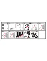
Figure 2
Figure 6
Figure 4
Figure 3
Figure 5
1. While looking through the eyepiece rotate the Vernier scale adjustment knob (figure 4) until the two half-shadows
are at the same dark intensity as the center section (figure 5).
2. The angle of rotation can now be read on the Vernier scales (figure 6) through the magnifying lenses located
on the eyepiece. A reading of 0° indicates that the sample is optically inactive.