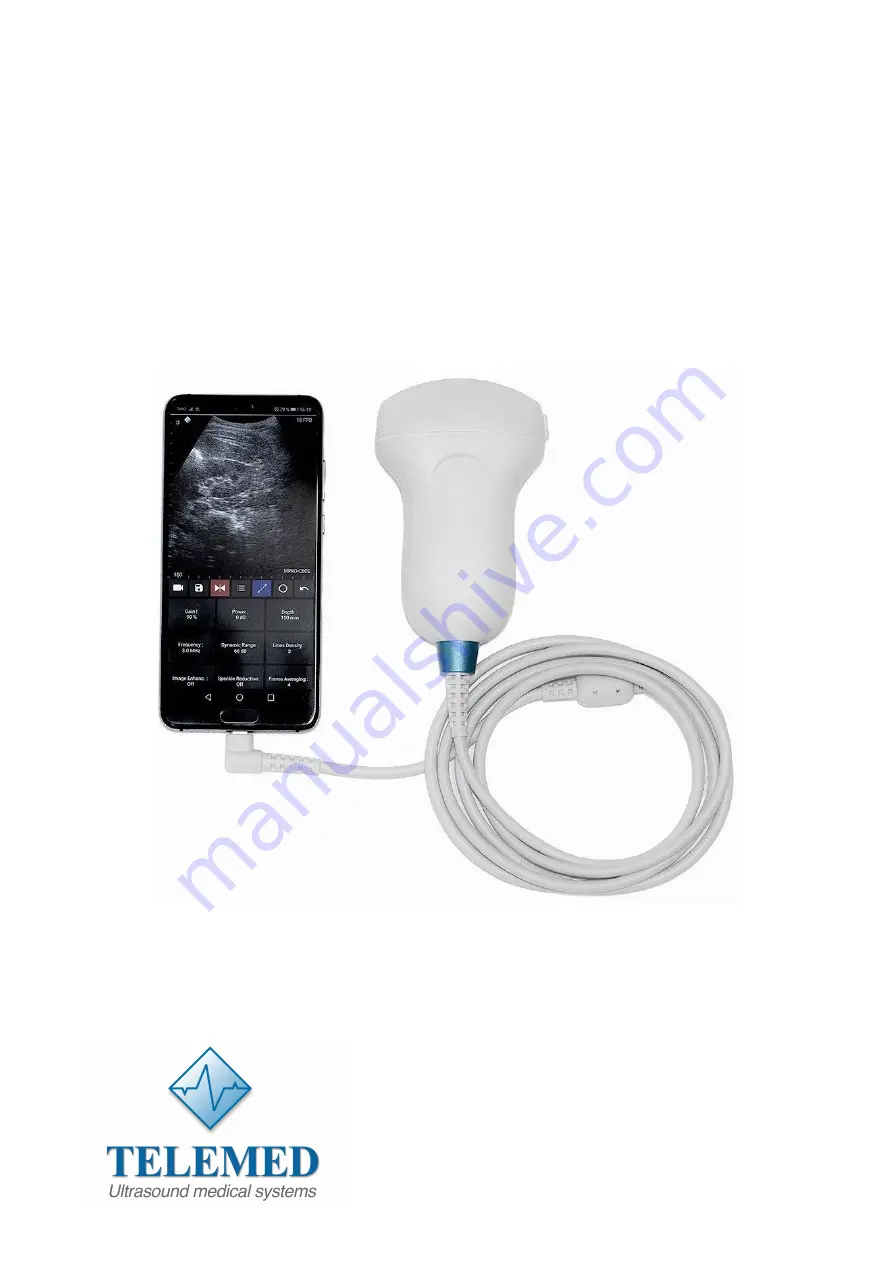
MicrUs and MicrUs Pro Series Ultrasound Systems
Echo Wave A Software
User Manual
TELEMED
Medical Systems
Italy
UAB Telemed
Lithuania
www.telemedultrasound.com/?lang=en
[email protected]
_
www.telemed.lt
Page 1: ...Ultrasound Systems Echo Wave A Software User Manual TELEMED Medical Systems Italy UAB Telemed Lithuania www telemedultrasound com lang en info telemedultrasound com support telemedultrasound com _www telemed lt _support telemed lt ...
Page 2: ... 3 Dynamic Range 12 7 1 4 Power 12 7 1 5 Gain 13 7 1 6 Change Scan Direction 13 7 1 7 Frequency 14 7 1 8 Frame Averaging 14 7 1 9 Rejection 14 7 1 10 Image Enhancement 15 7 1 11 Speckle Reduction 15 7 1 12 Lines Density 16 7 1 13 Map 16 7 1 14 Gamma Brightness Contrast 16 7 1 15 Negative 17 7 1 16 Zoom 17 7 1 17 TGC Time Gain Compensation 18 8 Measurements and Calculations 19 8 1 B Mode Measuremen...
Page 3: ...tware screenshots shown may look slightly different from the actual software user interface because user interface may depend on the used operating system adjustments In this document to refer to Echo Wave A ultrasound software may be used term ultrasound software or simply software Before using this software you must be trained in clinical procedures for conducting ultrasound examinations This gu...
Page 4: ...e phone or tablet computer and unlock the screen 2 Click Echo Wave A icon The icon may be not on first but on second or other page or on one of the pages of installed applications so you may need to sweep through pages to find it 3 After click you should see ultrasound software user interface 2 2 System Shutdown To exit Echo Wave A software click Back button of the operating system or phone tablet...
Page 5: ...User Interface Structure 3 1 Main Window 1 2 10 3 4 5 11 6 12 7 8 9 13 Num Description 1 Scan direction marker that indicates at what side is first probe element 2 Remaining phone battery indicator 3 Zero depth probe surface tick mark 4 Depth scale line 5 TGC Time Gain Compensation curve ...
Page 6: ...changed either continuously during movement sweep action e g Gain value or only when finger is released e g Depth value Required mouse movement type can be determined by observing hint image or at the right of parameter name For different scanning parameters are used different controls because different parameters have different number of values and require different time for programming of ultras...
Page 7: ...isplayed 6 Perform any measurements and calculations if required 7 Save ultrasound image 8 Press the Freeze Run button to restart the ultrasound scanning Ultrasound scanning freeze run state is indicated by the color of button and scan direction marker While scanning colors are as follows When scanning is stopped colors are as follows 9 Optionally repeat steps 1 8 to scan more ultrasound images De...
Page 8: ... frozen by clicking Freeze Run button and perform the following actions 1 Open the presets list by clicking on the Settings button then Presets tab 2 In the appeared list click on preset name in order to apply it If the number of available presets is large presets list can be scrolled by doing vertical sweeps with finger over opened presets list 3 Optionally repeat steps 1 2 to use another preset ...
Page 9: ...er preset name using on screen keyboard and click Save Preset button Presets are saved to folder Internal Storage Documents EchoWaveA Presets To delete ultrasound scanning preset please perform the following actions 1 Open the presets list by clicking on the Settings button and then Presets tab 2 Click preset deletion button 3 Click Yes when asked if you want to delete preset Please note that some...
Page 10: ...erform the following actions 1 Open the Settings window by clicking on the Settings button then click Patient button at the top 2 Click textbox after Patient ID 3 Enter patient identifier 4 Close keyboard and Settings window by clicking Back buttons Patient identifier if was entered appears in file names of saved image and video files ...
Page 11: ...y increasing the resolution of specific areas Always try to adjust focuses so that focusing markers are at the center of the anatomical structure which is of most interest Values The available list of focusing depth values depends on the probe used Focusing depth measurement units used millimeters mm Adjustment To change focusing depth wait while scanning controls will become inactive and then do ...
Page 12: ... every observation should be started using the maximum possible dynamic range since it provides you with the most complete diagnostic information Narrowing or reducing the dynamic range leads to the echo image displaying more contrast Dynamic Range is useful for optimizing tissue texture for different anatomical structures Values The values available depend on the type of probe used Measurement un...
Page 13: ...are filled in Gain adjustment may brighten or darken the ultrasound image if sufficient echo information is generated Values Gain values available are usually found in the interval 10 100 Measurement units used percentages Adjustment Click Gain button and do sweeps on ultrasound image Relations to other controls After adjusting the Gain you may need to adjust the Power level If you increase the ga...
Page 14: ...8 Frame Averaging Description Frame averaging is an image processing technique that allows you to obtain smoother softer images and reduces ultrasound image noise by averaging several sequential ultrasound image frames together Use higher frame averaging values to obtain smoother images Values The values available may differ depending on the type of probe but the following values are usually avail...
Page 15: ... enhancement methods available Off means that image enhancement is not used Adjustment Click Image Enhanc button and do sweeps on ultrasound image Relations to other controls Enabled image enhancement may reduce frame rate If your want higher frame rates you may need to turn off the image enhancement 7 1 11 Speckle Reduction Description Speckle reduction is an image enhancement technique that redu...
Page 16: ...ltrasound image Gamma 7 1 14 Gamma Brightness Contrast Description The software allows you to adjust the ultrasound image palette using gamma brightness and contrast controls Gamma adjustment changes the gray scale values of the ultrasound image in a non linear manner by raising the normalized values to the power of the chosen gamma value To make the entire ultrasound image appear lighter you can ...
Page 17: ...erform zoom in or zoom out of the B mode ultrasound image click Zoom button and do sweeps on ultrasound image Zooming also can be performed by touching B mode image with two fingers simultaneously and expanding contracting them When the image is zoomed and scanning controls are not active the image can be shifted by doing sweeps to any direction somewhere in the center of B mode image Default zoom...
Page 18: ...o the current scanning depth Small circular markers on this curve correspond to the TGC control points Values TGC values available are in the interval 0 100 in software they are not shown Measurement units used percentages Adjustment When scanning controls are not active press and hold for several seconds on ultrasound image at any depth at the right of depth scale line to show TGC curve Then whil...
Page 19: ...is do operation that in the future will be called press move release 3 Press move release on the ultrasound image to set second point and finish measurement 4 Optionally perform another measurement of the same type by repeating steps 2 3 Measurement object and result At first endpoint is displayed measurement number 1 and measurement result 48 6 mm Please note that when measurement operation is in...
Page 20: ...urement result 17 26 cm2 area P 154 5 mm circumference and V 43 236 cm3 volume Please note that for ellipse long axis first axis are marked two endpoint markers while for short axis second axis is marked only one endpoint 8 2 Deleting Performed Measurements To delete measurement click Delete Undo button Measurements are deleted one by one starting from last done measurement ...
Page 21: ...op button Video files in MP4 format are saved to Internal Storage Movies If video recording is not stopped manually the software will stop recording after 1000 frames Please note that writing to internal storage is done while recording so this may slow down ultrasound scanning Saved images and videos can be viewed using Android Gallery when ultrasound scanning software is closed and any viewer tha...
Page 22: ...vice supports this feature Freeze on software startup If turned on freezes ultrasound scanning on software startup Otherwise after software startup ultrasound scanning is running On freeze power saving If this option is turned on and is clicked button to freeze ultrasound scanning software turns off power of ultrasound device After clicking button to run ultrasound scanning device is powered on Th...
Page 23: ...ller value means smaller file size but also worse image quality MP4 video recording bitrate This option allows to adjust the quality of recorded MP4 video files Larger value means better quality but larger file size Smaller value means smaller file size but also worse quality Screen orientation This option allows to fix screen orientation and software layout in portrait or landscape position Portr...
Page 24: ...t button at the top In this window is also shown ultrasound device name and serial number By clicking buttons in this window you can view software user manual reference manual readme file software operation log file For viewing user manual and reference manual mobile device must have installed software that is registered to open PDF files For viewing readme and log file is required software that i...
Page 25: ...e do the following 1 Make software screenshot check user manual of your phone tablet how to do this 2 Copy software log file log txt from Internal Storage Documents EchoWaveA 3 Write down serial numbers of used ultrasound scanner and probe 4 Write down phone tablet model 5 Describe the problem and steps to reproduce it 6 Send all above mentioned information to support telemed lt 14 References 1 Ec...