background image


Revised 03/25/2020 


20 


7.) 

The MF knobs do not move the diffraction pattern


Occasionally, the microscope will stop auto-assigning the multifunction knobs to 
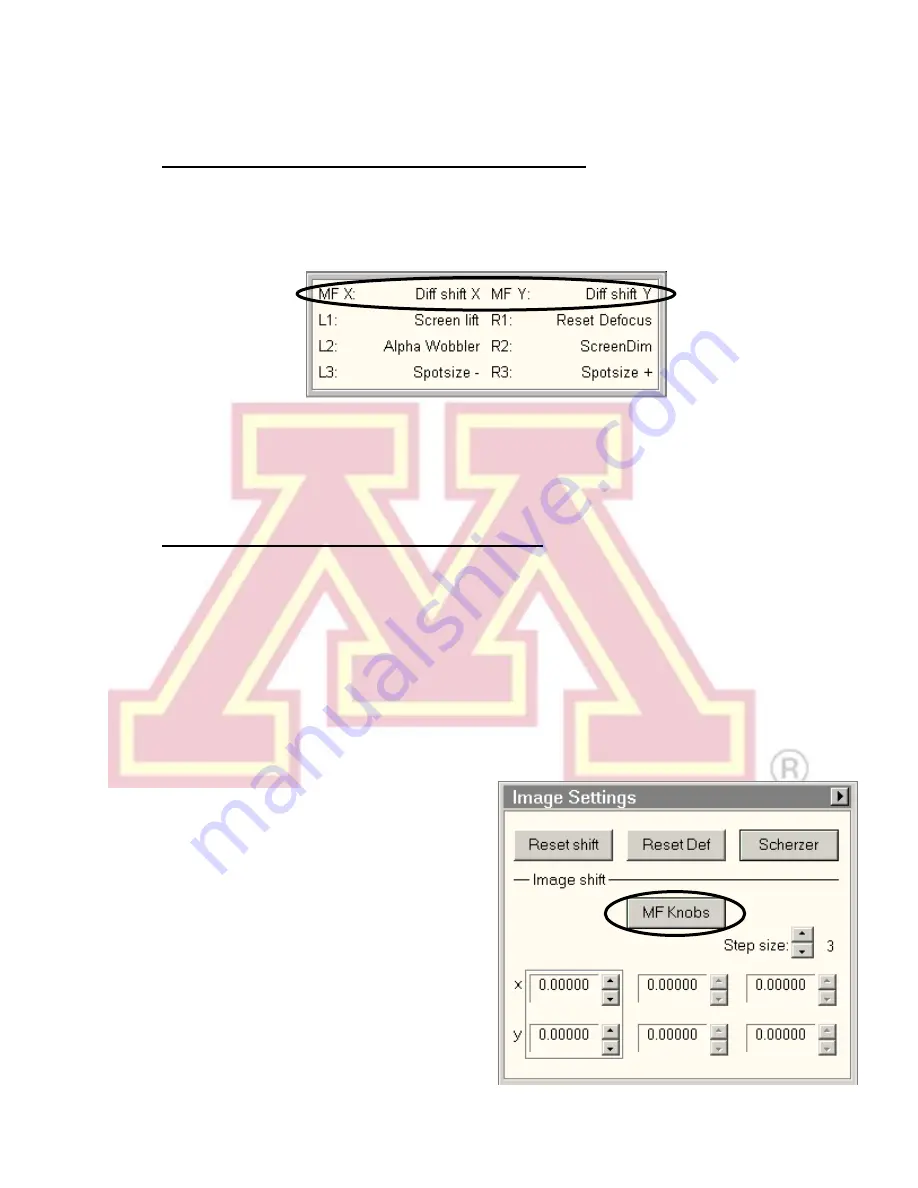
diffraction shift when entering diffraction mode. Check the lower-left status box to 
verify that the MF knob assignments read “Diff shift”. 


If the MF knobs are assigned to a stigmator, this will override the diffraction shift. 
Turn the stigmator off to return to diffraction shift. Otherwise, you can manually 
assign it 

by right clicking in the “MF X” box and selecting “Diff shift X”. Be sure to 

right click again and select “None” when finished adjusting the pattern. 

8.) 

Large amounts of specimen drift are present


There are several possible causes of specimen drift. The following may reduce it: 

a.) Lightly  tapping  on  the  end of  the  sample  holder  rod  can  cause  it  to  settle  in 

the goniometer, reducing drift. 

b.) Move to a different area of the sample. Damaged regions of a support film will 

often  move  as  they  are  subjected  to  the  electron  beam.  In  this  case,  the 
sample  itself  is  moving,  and  viewing  a  more  stable  region  may  reduce  or 
remove the apparent drift. 

c.)  Avoid  moving  the  stage,  when 

possible. Every stage movement will 
require  a  settling  time  once  it 
completes.  Use  the  image  shift 
function  to  make  small  adjustments 
to the view without moving the stage. 

Image  shift  can  be  assigned  to  the 
multifunction  knobs  by  locating  the 
“Image  Settings”  control  panel 
(usually  found  in  the  “Camera” 
workspace tab) and clicki

ng the “MF 

Knobs”  button.  Click  again  to  return 

Summary of Contents for T12

Page 1: ...is not lit press the button to light it and proceed normally but make a note in the instrument log 2 Log in with the username and password created during training If the vacuum logger is running close it and log off vacuumuser first 3 Sign in to the CharFac reservation system and the paper logbook 4 Launch the Tecnai User Interface and Digital Micrograph programs 5 Check that the microscope status...

Page 2: ...en by selecting it from the box in the bottom right corner of the screen Check that IGP1 Gun Col reads 6 log units P3 indicates the camera vacuum level It should read 35 P1 indicates the pressure in the buffer tank and P2 the pressure in the backing pump line This dialogue box is used to select the vacuum overview or any of several other information or setup tabs ...

Page 3: ... Continue in 20 kV increments waiting 1 2 min at each If the emission current remains high 1 2 µA return to the previous step for several minutes Note The HT may switch off and become disabled when reaching 40 kV If this occurs simply press the main HT console button to re enable it see Section I click the High Tension again at 40 kV and continue III Specimen Loading and Holder Insertion Removal N...

Page 4: ...il it stops This rotation moves the guide pin see steps 3 and 4 approximately from the 12 o clock position to 5 o clock d Gently while keeping pressure on the goniometer pull the sample holder back to break the airlock vacuum This will require a small amount of force e Remove the holder straight back out of the column while being careful not to scrape it along the inside of the airlock f Be carefu...

Page 5: ... and gently raise the clamp straight up until it stops d Place the specimen grid into the recess at the end of the holder e Gently lower the clamp straight down to hold the grid securely Return the tool to the base of the holder stand f Retract the holder slightly and turn it upside down Tap the back end several times then turn the holder upright and check that the grid has not moved movement sugg...

Page 6: ...mpu stage will go on Do not move the holder while the red stage LED is lit c The pumping time remaining will be visible in the Vacuum Overview window d Select the specimen holder type Single Tilt from the box in the interface Be sure to click the button to confirm the selection e When the pump times ends status reads COL VALVES and the red stage LED goes out support the purple goniometer surface w...

Page 7: ...n Step setting controls the bias voltage on the Wehnelt cylinder A higher step value decreases the bias This produces more emission current but increases the energy spread and source size of the beam V Alignment Note Align the microscope from the top gun down Press F1 at any time for online help with an alignment Begin with the objective and SA apertures removed Leave a condenser aperture inserted...

Page 8: ...agnification RC Magnification in the 10 60 kx range and set Spot size to 3 b Center the beam using the beam shift LC trackball and spread the beam LC Intensity clockwise from crossover to 3 4 the size of the screen Note If the beam is very asymmetrical roughly adjust the condenser stigmation see step 5 c Select Gun Tilt from the Direct Alignments panel in the Tune workspace d Adjust the gun tilt u...

Page 9: ...sizes 3 and 9 g Press Done in Direct Alignments h If the gun shift must be adjusted a substantial amount and it results in a significant change in the measured exposure time at a given spot size re check the gun tilt alignment 4 Centering the Condenser Aperture a Select the desired condenser aperture 4 is largest 1 is smallest using the large outer knob on the aperture b Converge the beam to cross...

Page 10: ... Adjustment Note all alignments beyond this point depend on the objective focus To ensure proper alignment both the specimen and objective lens focus must lie on the eucentric plane of the microscope This step will position the specimen a Find a point of interest on the specimen using the stage trackball RC b Activate the Alpha wobbler LC button L2 by default The stage will begin rocking through a...

Page 11: ...ents f Repeat steps b e for Beam tilt pp Y 8 Beam Shift a Converge the beam to crossover LC Intensity and center it using the LC trackball b Select Beam shift from Direct Alignments and use the MF knobs to re center the beam on the screen c Press Done in Direct Alignments 9 Rotation Centering Note Rotation centering is the most important alignment for high resolution work on this microscope It sho...

Page 12: ...central diffraction spot using the MF knobs b Select and insert the desired objective aperture see aperture figure in step 4 The aperture should be visible in the diffraction plane c Center the aperture around the central spot aperture X and Y knobs d Deselect Diffraction to return to imaging mode Note If the aperture is not visible in the diffraction pattern do not randomly adjust the aperture as...

Page 13: ... tune the stigmator settings e Click None in the Stigmator window or press the LC Stigmator button Note Objective astigmatism should be checked periodically throughout a session It can be changed by changes in magnification mode spot size Z height of the sample or objective aperture size or position Note Each stigmator has three registers available Clicking on a register selects among them These c...

Page 14: ...the beam can reach the camera d There are three camera modes Search is a high refresh rate lower resolution mode used for viewing and focusing on the specimen in real time Preview is a slower higher resolution mode that uses half the CCD area to preview the final acquired image quality Acquire is used to a capture a single image from the whole CCD detector Note The parameters for any of these sett...

Page 15: ...u start typing your username it should jump to it You can drag files from the T12 PC to the right hand window to transfer them to your storage directory If you use multiple instruments you may want to make a T12 directory here e After transfer close WinSCP and shutdown normally f See addendum for instructions on remote file access 3 Dark and Gain References If consistent artifacts are present in a...

Page 16: ...ndenser aperture inserted in out out b Leave the column valves closed c Place the viewing screen down cover the window with the rubber mat d Switch the filament off Filament button in the Filament panel e Leave the magnification in the SA range preferably 2400 This is essential to maintain stable objective lens current and prevent thermal drift for the next user f Remove the sample holder be sure ...

Page 17: ...um monitor username vacuumuser password vacuum c The Vacuum Logger software should start automatically Click Log Start to begin logging the vacuum levels Create a new file using today s date mm dd yyyy as the file name include zeros in the day month VIII Troubleshooting 1 No beam is visible There are several possible causes for no beam being visible Try the following steps until a beam can be foun...

Page 18: ...button then click apply If desired alignments for other systems modes of the microscope can also be loaded by moving the appropriate labels to the Selected list and pressing apply 2 One of the control pads or z axis or tilt buttons is unresponsive Re initialize the pad by unplugging the USB cable from the back of the PC and re inserting it The right plug connects to the right panel left to the lef...

Page 19: ...it should be visible in the back focal plane when you return to SA mode diffraction 4 Images contain black specks ghosts or other artifacts Make sure that both dark and gain references are being applied to the image Collect new references if necessary see section VI step 3 on pages 15 16 5 The stage or beam shift trackball is unresponsive If other controls on the same pad function normally then tr...

Page 20: ...re present There are several possible causes of specimen drift The following may reduce it a Lightly tapping on the end of the sample holder rod can cause it to settle in the goniometer reducing drift b Move to a different area of the sample Damaged regions of a support film will often move as they are subjected to the electron beam In this case the sample itself is moving and viewing a more stabl...

Page 21: ...he door open talking or large groups present in the instrument room will inevitably produce thermal and acoustic variations that will result in specimen drift Avoid these situations whenever possible 9 Other problems If the software is behaving unexpectedly you may close and restart the user interface and Digital Micrograph Logging out and back in may also resolve some problems Do not restart the ...

Page 22: ...each instrument is FEI T12 Shepherd TEM T12 umn edu FEI FEGTEM Shepherd F30 TEM F30_Shepherd umn edu FEI Cryo FEGTEM NHH NHH F30 TEM F30_NHH umn edu Messages sent to the mail list will be distributed to everyone on the list Some uses for the mailing list include To subscribe unsubscribe Email addresses at umn edu can subscribe automatically through listserv umn edu To subscribe non umn edu address...

Reviews: