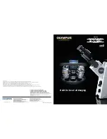
©
Home Training Tools Ltd. 2005
Page 3 of 7
Visit us at www.homesciencetools.com
4.
Zoom knob:
This knob is used to change
magnification. It allows you to “zoom” from
10x to 40x.
5.
Objective turret:
This turret contains the
lenses closest to the specimen. The objective
lenses have magnification between 1x and 4x
(providing a total magnification of 10x-40x
when multiplied with the 10x of the
eyepieces).
6.
Locking knob:
The binocular head is
mounted on a post and can be raised,
lowered, or turned around by loosening the
locking knob on the back of the post.
7.
Top illuminator:
This bulb-holder holds the
10-watt halogen bulb that shines down on the
specimen. Use this light when your specimen
is opaque or solid (when light cannot pass
through it from below).
8.
Focus knob:
This knob is used to raise or
lower the objective lens until the image is in
focus.
9.
Stage:
The stage is the platform that supports
the specimen below the objective lens.
10.
Head stop:
This sets the lowest position the
head can drop. For normal use it can be left
in the lowest position. If you are examining
tall specimens, adjust the ring so that the
head cannot hit the specimen.
11.
Stage plate screw:
The stage plate can be
removed and changed by loosening this
screw.
12.
Stage clips:
These clips can be used to hold
thin specimens in place.
13.
Stage plate:
This microscope comes with two
stage plates. The glass plate is used with
bottom lighting, and the reversible black/white
plate is used with top lighting to help you get
the best contrast.
14.
Bottom illuminator:
Another 10-watt halogen
bulb is located beneath the stage plate. Use
this light for translucent specimens.
15.
Illuminator control:
This allows you to
choose three different light settings: top
lighting, bottom lighting, or top and bottom
together.
Operating Procedure
Now that you have an overview of what each
component of your microscope is for, you can
follow this step-by-step procedure to help you get
started using it.
Getting Started
1.
Set your microscope on a tabletop or other flat
sturdy surface where you will have plenty of
room to work. Plug the microscope’s power
cord into an outlet, making sure that the
excess cord is out of the way so no one can
trip over it or pull it off of the table.
2.
Flip the switch to turn on your microscope's
light source. Use top lighting for opaque
specimens and bottom lighting for translucent
specimens. Some specimens have both
opaque and translucent parts. For these use
top and bottom lighting together.
Warning:
The top light can get very hot. Use care
touching the top light housing during use.
3.
Center a specimen on the stage plate. If you
are using top lighting, insert the reversible
black/white stage plate (use the dark side if
the specimen is light colored). To change or
reverse the plate, loosen the stage plate
screw until you are able to pop the plate out.
Turn the plate over and tighten the screw to
lock it in place.
4.
If your specimen is thin and flat, or if its edges
curl up easily, use the stage clips to hold it in
place. To do this, pull up the pointed end of
one stage clip and slide it over one end of the
specimen, then do the same with the stage
clip on the other side. If your specimen is
larger than the stage plate, turn the stage
clips out so that they hang off the stage; this
will give you more room to work.
5.
You may need to adjust the height of the head
in order to find a good working distance
between the specimen and the objective lens.
Do this by loosening the locking knob, moving
the head to the appropriate position, and
tightening the locking knob.
6.
Turn the zoom knob away from you until the
microscope is on its lowest power.
7.
Slowly turn the focus knob until the specimen
comes into view. Once you can see the
outline of the specimen, turn the knob even
more slowly until it is focused as sharply as
possible. Once you have focused your
specimen, you can move it around to see
other parts of it. You may need to refocus
slightly on each new area.
Note
: with this
microscope you will often be viewing three-
dimensional specimens that have many