

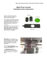


3000-LED SLIDER PHASE CONTRAST INSTRUCTIONS
PHASE CONTRAST
MICROSCOPE
The normal microscopic object is seen
because it has regions of varying density. In
normal brightfield illumination a completely
transparent specimen is very difficult to
observe in detail because all areas of the
specimen are equally dense. Darkfield
illumination displays border effects in
completely transparent specimens due to
edge scattering and diffraction of light.
Polarized light is useful when transparent
specimens have directional or crystalline
properties.
Phase contrast microscopy is a type of
illumination system to observe transparent
media. This form of illumination is utilized
extensively in the study of transparent living
cells without the need for staining or fixing
while being able to obtain good image
contrast. The light from phase contrast
illumination arrives at the user’s eyes at ½
the normal wavelength. This light altering
system produces a visible image of an
otherwise invisible, transparent specimen.
The optical light path necessary for phase
contrast is shown in Figure 1. A clear annulus
in the focal plane of the condenser is imaged
at infinity by the condenser and then re-
imaged by the objective in its rear focal
plane. The undiffracted light passes through
this image. It is reduced in intensity and given
a one-quarter wave phase shift by means of
an annular phase pattern in the rear focal
plane of the objective. These two changes in
the undiffracted portion of the beam simulate
the phase and intensity distribution which
would be present in the objective focal plane
if the specimen had density variations rather
than refractive index variations. As a result,
the image formed by the beam interfering
with the diffracted beam simulates that of a
specimen having density variations.
IMAGE FORMATION
BY PHASE CONTRAST
An annular aperture in the diaphragm placed in the focal
plane of the substage condenser controls the illumination
of the specimen. The aperture is imaged by the
condenser and objective at the rear focal plane or at the
exit pupil of the objective. A phase shifting element, or
phase plate, is placed in the image plane. Light passing
through the phase altering pattern acquires a ¼ wave
length advance over that diffracted by the object structure
and passes through that region of the phase plate not
covered by the altering pattern. The resultant interference
effects of the two portions of light form the final image.
Altered phase relations in the illumination rays, induced
by otherwise invisible elements in the specimen, are
translated into brightness differences by the phase
altering plate.
Summary of Contents for 3000-LED series
Page 2: ......