
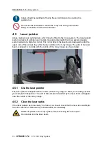


Getting
started
XT V 160 X-ray System
XTM0011-F2
43
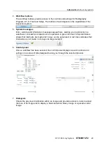
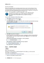
Save the current image to disk
You can save captured images that you have enhanced or analysed.
Click the down arrow next to
to change the save image options.
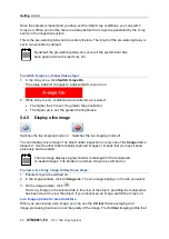
To save an image, follow these steps:
1. On the image toolbar, click
.
The
Save As
dialog box opens.
2. Browse to and select the folder where you want to save the image.
3. Type a name for the image.
4. Select the image file type. You can save the image as a TIFF, Bitmap or JPEG.
5. Click
Save
.
If you save the image in colour, with annotations, or not in a TIFF
format, you will not be able to load this image back into Inspect-X.
Depending on the setting in the
Imaging Properties
, saving the image
again will either overwrite the original or open the
Save As
dialog box,
allowing you to save the image with a different filename.
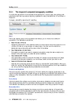
5.5
Computed tomography
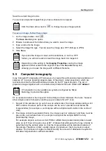
X-ray Computed Tomography (CT) allows you to create three-dimensional representations of
samples. CT involves acquiring angular views of the object, called projections, which are
reconstructed into the three-dimensional volume by the CT Agent program. The set of
projection images together with a reconstruction parameter files are called a
CT dataset
.
CT acquisition is only available on systems configured by Nikon
Metrology to provide this option.
CT is covered in detail in the
Inspect-X Online Help and User Manuals
. There are, however,
a few simple rules to follow when setting up your sample for a CT scan.
1. No part of the sample can go out of view on either side of the image window during a full
360° rotation. However, parts of the sample can be out of view above and below the
image window (for example, a vertical pipe with an elliptical weld visible in the centre of
view).
2. The image cannot be saturated (that is, the image must not 'white out' and there must be
grey scale, even where there is no sample in view) but the sample MUST be fully
penetrated by X-rays.
For example, objects such as a small PCB or a BGA have greater density when viewed
edge on at 0° than in the plan view at 90°. It follows that while using the X-ray intensity to
penetrate fully the sample edge on, that the image window will be saturated outside of
the sample. Therefore, a filter, usually copper, is placed over the X-ray source output
window. The thickness of copper is determined by the amount of attenuation required to
still allow full penetration, without saturation.
Содержание XT V 160
Страница 1: ...X Tek X ray and CT Inspection XT V 160 X ray System Operator Manual XTM0011 F2 ...
Страница 2: ......
Страница 4: ......
Страница 6: ......
Страница 8: ......
Страница 22: ......
Страница 26: ......
Страница 32: ......
Страница 83: ...Maintenance XT V 160 X ray System XTM0011 F2 73 ...
Страница 90: ...Troubleshooting 80 XTM0011 F2 XT V 160 X ray System ...
Страница 93: ......


