


Electrooculography (EOG)
User Manual
4 of 17
1.
General Information
1.1.
General Description
The biosignalsplux Electrooculography (EOG) sensor has been specially designed for seamless EOG
data acquisition. Either used by itself or in combination with an eye tracker, our sensor can provide
additional insight into your subjects’ eye gaze patterns.
The bipolar configuration, with two
measurement electrodes, detects the electrical potentials in the specific temporal or facial region of
choice, with respect to a reference electrode (placed in an area of low bioelectrical activity). The
resulting signal is the amplified difference between these two leads, eliminating the common
unwanted signals. Its convenient form factor enables a discrete application in the typical EOG
electrode placement locations.
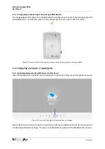
Figure 1: biosignalsplux EOG sensor (standard version)
1.2.
Typical Unfiltered Sensor Output
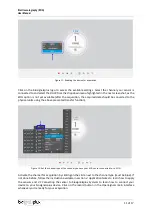
Figure 2 shows a typical unfiltered Electrooculography sensor output acquired while blinking. The raw
digital sensor values received from the biosignalsplux device ranged between 0 and 2
n
-1 (n=sampling
resolution) were converted into the original unit of measurement of this sensor (mV) using the transfer
function found in section Transfer Function (Conversion Formula).
Figure 2: Typical unfiltered sensor output (while blinking and the sensors positioned above and below the eye).