Posterior Segment
2660021169012 Rev. A 2017-12
CIRRUS HD-OCT User Manual
8-30
2. The normative database consisted of a population with a limited range of spherical
error (-12 D to +6 D) and axial length (22 to 28 mm). Subjects with strongly myopic or
hyperopic eyes may have a different distribution of measured RNFL thickness values,
and may tend to flag more often than subjects who fall within the range of the
population used to create the normative database.
There is a wide variation in RNFL bundle anatomical distribution among the normal
population. A person with split-bundle anatomy or a person with a very tilted RNFL bundle
pattern may show a deviation from normal anatomy without indicating that this person
has lost RNFL.
Changing RNFL Calculation Circle Placement
Changing the placement of the RNFL Calculation Circle changes the Deviation from Normal
Map as well as the optic disc parameter calculations, since each super-pixel in the scanned
area is defined relative to the center of the Calculation Circle. Super-pixel positions in the
normative data are defined relative to a fixed center based on the age-matched normative
samples. Therefore, when you change the position of the Calculation Circle, you change
the specific super-pixel in the normative data against which each super-pixel in the exam
data is compared.
NOTE: When the temporal RNFL is very thin or entirely absent, the RNFL algorithm may
show an artificial thickening of the RNFL in this area. If the temporal RNFL appears thicker
than normal, examine the algorithm lines as displayed on the extracted circular B-scan to
determine if the algorithm has correctly identified the RNFL.
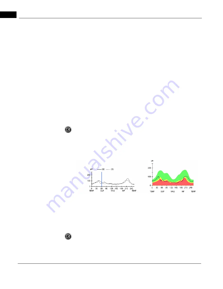
TSNIT Thickness Profiles
The TSNIT Thickness Profiles (TSNIT stands for Temporal, Superior, Nasal, Inferior, Temporal)
display thickness at each A-scan location along the Calculation Circle and include as a
backdrop the white-green-yellow-red color code based on the age-matched RNFL
normative data. The profile shows left and right eye RNFL thickness together, to enable
comparison of symmetry in specific regions. Drag the blue vertical line in the OU profile to
select the current A-scan sample from among the 256 samples.
NOTE: You cannot select every specific A-scan sample by dragging the vertical blue line. To
select an individual A-scan, click (and release) the vertical blue line, then hold down Ctrl
and press the left or right arrow key.
Summary of Contents for CIRRUS HD-OCT 500
Page 1: ...2660021156446 B2660021156446 B CIRRUS HD OCT User Manual Models 500 5000 ...
Page 32: ...User Documentation 2660021169012 Rev A 2017 12 CIRRUS HD OCT User Manual 2 6 ...
Page 44: ...Software 2660021169012 Rev A 2017 12 CIRRUS HD OCT User Manual 3 12 ...
Page 58: ...User Login Logout 2660021169012 Rev A 2017 12 CIRRUS HD OCT User Manual 4 14 ...
Page 72: ...Patient Preparation 2660021169012 Rev A 2017 12 CIRRUS HD OCT User Manual 5 14 ...
Page 110: ...Tracking and Repeat Scans 2660021169012 Rev A 2017 12 CIRRUS HD OCT User Manual 6 38 ...
Page 122: ...Criteria for Image Acceptance 2660021169012 Rev A 2017 12 CIRRUS HD OCT User Manual 7 12 ...
Page 222: ...Overview 2660021169012 Rev A 2017 12 CIRRUS HD OCT User Manual 9 28 ...
Page 256: ...Log Files 2660021169012 Rev A 2017 12 CIRRUS HD OCT User Manual 11 18 ...
Page 308: ...Appendix 2660021169012 Rev A 2017 12 CIRRUS HD OCT User Manual A 34 ...
Page 350: ...CIRRUS HD OCT User Manual 2660021169012 Rev A 2017 12 I 8 ...
Page 351: ...CIRRUS HD OCT User Manual 2660021169012 Rev A 2017 12 ...


