background image

101

SPECIFICATIONS AND PERFORMANCE

SPECIFICATIONS AND PERFORMANCE

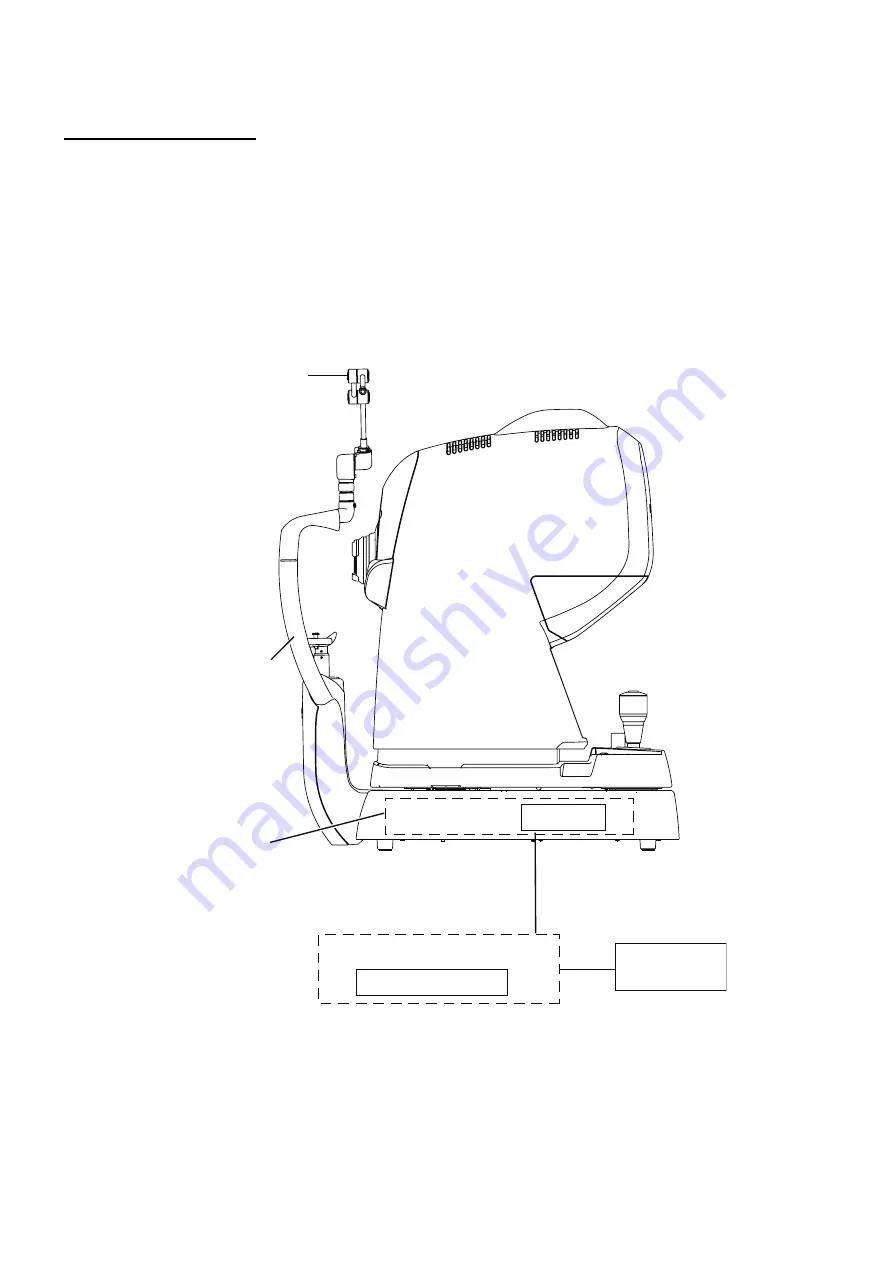
SYSTEM DIAGRAM

This instrument is composed of the following units.

 Instrument body (main unit, chin-rest unit and power supply unit)

 External fixation target

 Power cord

 LAN cable

 Personal computer (including the personal computer main unit, monitor, mouse and keyboard)

 Insulation transformer

 Analysis software

LAN terminal

Power supply unit

Chin-rest unit

Main unit

External fixation target

LAN cable

(Analysis software)

Personal computer

Insulation 
transformer

Summary of Contents for DRI OCT-1 Triton

Page 1: ...USER MANUAL 3D OPTICAL COHERENCE TOMOGRAPHY DRI OCT 1 Model Triton DRI OCT 1 Model Triton plus...

Page 2: ......

Page 3: ...ting the anterior segment attachment kit which is an optional accessory you can observe photograph and record the tomogram of anterior segment After photographing the images and tomograms of fundus an...

Page 4: ...all terms and conditions of IPA Font License Agreement v1 0 shall be deemed to be accepted and agreed by you if you use DRI OCT 1 Model Triton For IPA Font License v1 0 refer to the following URL htt...

Page 5: ...STATE 32 BASIC OPERATIONS 33 FLOW OF OPERATION 33 PREPARATION FOR PHOTOGRAPHY 34 FUNDUS TOMOGRAPHY 35 DELETING DATA 48 SAVING DATA 48 PRINTING DATA 48 HOW TO FINISH 49 OBJECTIVE OPERATIONS 50 COLOR FU...

Page 6: ...8 STORAGE USAGE PERIOD 108 ENVIRONMENTAL CONDITIONS FOR PACKAGING IN STORAGE 108 ENVIRONMENTAL CONDITIONS FOR PACKAGING IN TRANSPORTATION 108 ELECTRIC RATING 109 DIMENSIONS AND WEIGHT 109 SYSTEM CLASS...

Page 7: ...rdous situation which if not avoided may result in minor or moderate injury NOTE Useful functions to know Paying attention to these will prevent the noted problems Symbol IEC ISO Publication Descripti...

Page 8: ...accessories sold with this product will expose you to lead a chemical known to the State of California to cause birth detects or other reproductive harm Washhandsafterhandling Preventing Electric Shoc...

Page 9: ...ent Request service from an authorized Topcon distributor Do not put any substance over the vent on the top surface of the power supply unit If the vent is cov ered the temperature of the power supply...

Page 10: ...due to fire earthquakes actions by third persons and other accidents or damage due to negligence and misuse by the user and any use under unusual conditions TOPCON shall not take any responsibility f...

Page 11: ...not to bump the patient s eye or nose with the instrument when operat ing the touch panel PR CAUTION Pour viter une blessure du patient faire attention de ne pas heurter les yeux ou le nez du patient...

Page 12: ...ternal fixation target 1 Monitor cleaner 1 User manual Unpacking and assembly manual and Analysis software DVD 1 each Dust cover 1 Accessory case 1 Chinrest tissue pins 2 LAN cable 1 UNPACKINGANDASSEM...

Page 13: ...t Chinrest unit External fixation target Diopter compensation lens selector Focusing knob Base clamping knob Touch display External connection terminal External cover Photography button Control lever...

Page 14: ...button Starts photography Base clamping knob Locks and unlocks the base Small pupil diaphragm selector button Sets the small pupil diaphragm to ON OFF Split button Sets the split lines to ON OFF Exter...

Page 15: ...DISPLAY NOTE Operate the touch display by your fingers Don t use any sharp tool such as a ball point pen The touch display may be damaged to cause an incorrect operation Tap To select any relevant ite...

Page 16: ...s the capture icons Follow up button Each time you press this button Follow up ON button is orange and Follow up OFF button is black is changed each other In case of ON Follow up photography is done I...

Page 17: ...n the relevant data does not exist One eye data of the relevant data does not exist Both eyes data of the relevant data does not exist NOTE Follow up photography function in DRI OCT 1 Model Triton is...

Page 18: ...gment live image area This area displays the live image of fundus or anterior segment and the graphic image of the scan pattern which is set on the selected capture icon with an interrupted line The r...

Page 19: ...hy button Displays the color fundus photography status ON OFF You can change ON OFF of color fundus photography FAF photography button Only in DRI OCT 1 Model Triton plus Displays the FAF photography...

Page 20: ...e Cross the arrow direction for the right eye is reversed for the left eye The yellow line is displayed for the scan pattern except Line For 3D the yellow line shows the scan range and for other scan...

Page 21: ...layed only for Line and 5 Line Cross scans Each time you touch these buttons the scan line is rotated by 1 in the arrow direction RESET button Press this button The changes for the position which were...

Page 22: ...green and yellow Displays the graphic image of the scan pattern which is set on the selected capture icon The green line shows the scan position and the arrow shows the scan advance direction The arro...

Page 23: ...he screen Refer to P 40 Tracking photography The display color of the scan pattern on the IR fundus image shows whether tracking is successful or not in Radial Line and 5 Line Cross scan When tracking...

Page 24: ...ogram Z lock position display bar Indicates the Z lock position changeable range By touching an optional position on the bar you can move the center position of the displayed tomogram to the touched p...

Page 25: ...dealer or TOPCON listed on the back cover Image Quality level meter Displays the Image Quality level with meter Live image Displays the live tomogram image Stereoscopic image Displays the image of th...

Page 26: ...this area In 3D scan the two images at both ends are regarded as the typical images and in Radial scan the two images vertical and horizontal are regarded so For each image an icon that indicates the...

Page 27: ...l bar at the right Page number Displays the current setting menu page s number in page order Icon layout setting area Sets the icons that will be displayed on the Capture icon selection screen which i...

Page 28: ...the icon is added to the left side or upper right corner of the capture icon Delete icon button Deletes the capture icon which is selected in the icon layout setting area After deleting the remaining...

Page 29: ...ame Displays the objective items of the parameters for the selected cap ture icon Item value selector button Displays the set value of each item Select the desired item OK button Validates the changed...

Page 30: ...age s number in page order Utility button Displays the objective items on the current setting menu page Select the desired item Page forward backward button Shifts to other pages in the setting menu I...

Page 31: ...instrument with the base clamping knob at the bottom To avoid injury or damage two people should be employed for sup porting the instrument from the underside Carrying by one person may result in pers...

Page 32: ...to the instrument body 3 Plug the power cord into the 3 pin plug AC receptacle WARNING To avoid fire and electric shock in case of leakage be sure to use a power supply equipped with a 3 pin plug AC r...

Page 33: ...of the instrument NOTE Use the external device complying with IEC60950 1 Do not connect any device that is not specified as a system For details about connecting the external devices contact your dea...

Page 34: ...on and focusing knob is not operated for a period of time the power save function stops power supply to the touch display illumination light source and photography light source 1 Tap the control panel...

Page 35: ...er P 34 Inputting the patient information Setting the patient P 37 Setting the illumination level P 40 Changing the diopter compensation lens P 41 Alignment and photography P 42 Transferring the data...

Page 36: ...the instrument to the personal computer in which the analysis software the acces sory of the instrument is installed 2 Turn on the personal computer 3 Start the analysis software 4 Enter the patient i...

Page 37: ...ter of the picture The patient s pupil is small This condition includes the case of When the small pupil diaphragm is set to ON The flash level is high Alignment is not adjusted properly To improve th...

Page 38: ...resolution Fixation position Myopia Mode Over Scan Count Line H Line V 9 0mm 1024H 1024V Macula OFF 128 5 Line Cross 6 0mm 1024 10 Macula OFF 16 Radial Dir 6 0mm 1024 12 Macula OFF 16 3D Macula 7 0 7...

Page 39: ...de corner of the patient s eye is level with the Canthus marker on the chinrest post Let the patient rest his her forehead on the forehead rest CAUTION To avoid electric shock do not touch the externa...

Page 40: ...e target is changed to the external fixation target Change to the desired picture position For the details of display refer to P 17 Changing by using the internal fixation target position move button...

Page 41: ...buttons around the image To use rotation which can be adjusted only for Line scan and 5 Line Cross scan perform the rotating adjustment with the rotation buttons clockwise counterclockwise indicated a...

Page 42: ...dus anterior segment live image area and the flash level increases Tap the and the flash level decreases When color fundus photography is set to OFF or when the instrument without the fundus photograp...

Page 43: ...ye has a strong myopia turn the diopter compensation lens selector and set it to When the patient s eye has a strong hyperopia turn the diopter compensation lens selector and set it to Compensation ra...

Page 44: ...turn the control lever right for upward movement and left for downward movement The vertical position mark indicates the standard for the instrument s vertical position CAUTION To avoid injury while m...

Page 45: ...f the fundus live image area 3 On the touch display bring the scale towards the patient s pupil and make sure that the pupil is larger than the scale NOTE Now hold the control lever upright which will...

Page 46: ...e Illumination level display on the touch display 7 Bring the base straight toward the patient side Two alignment bright spots for the working distance alignment and the split lines for the focal dist...

Page 47: ...focusing knob to change the split lines into one line If you cannot align the split lines into one line by turning the focusing knob change the diopter compensation lens Refer to Changing the diopter...

Page 48: ...nal fixation target Set the system to detect the retina position automatically and optimize the image output sensi tivity NOTE If the patient blinks frequently the auto functions do not work smoothly...

Page 49: ...sing the OCT pho tography is done and then the fundus photography is automatically done 14Each time you take a picture the preview screen appears 15Tap the photography mode button at the top on the sc...

Page 50: ...eleted When color fundus photography is set to OFF or when the instrument without the fundus photography function is used the color fundus photography image is changed to the IR fundus photography ima...

Page 51: ...nt the base from moving suddenly NOTE To prepare for next photography turn the control lever and move the instru ment body to the center position The vertical position mark indicates the standard for...

Page 52: ...n ter of the picture The patient s pupil is small This condition includes the case of When the small pupil diaphragm is set to ON The flash level is high Alignment is not adjusted properly To improve...

Page 53: ...Refer to Setting the illumination level on page 40 Setting the flash level Set the flash level by touching the Flash level display on the fundus anterior segment live image area Refer to Setting the f...

Page 54: ...ears on the fundus anterior segment live image area 5 The scale on the touch display moves to the alignment position corresponding to the picture position 6 Ask the patient to see the green light inte...

Page 55: ...right before the color fundus photography the automatic blink detection function stops the photography Start the procedure again from alignment and take a picture When the auto shoot function is OFF o...

Page 56: ...hotography Under the following photography conditions there is a bright spot on the cen ter of the picture The patient s pupil is small This condition includes the case of When the small pupil diaphra...

Page 57: ...2 Photography Setting on P 82 Setting up the patient and capturing Refer to Setting up the patient on page 37 NOTE In peripheral photography the automatic small pupil function does not work Fixation p...

Page 58: ...the flash level on page 40 Changing the diopter compensation lens Turn the diopter compensation lens selector to change the diopter compensation lens to 0 Refer to Changing the diopter compensation l...

Page 59: ...ient side and the fundus image appears on the fundus anterior segment live image area 5 The scale on the touch display moves to the alignment position corresponding to the picture position 6 Ask the p...

Page 60: ...base unit toward the patient in stereoscopic photography the focusing knob is not reset to the initial position When the auto focus function is OFF turn the focusing knob to change the split lines in...

Page 61: ...nction A picture of another eye is taken and the preview screen appears NOTE If the light intensity of the photographed image is not correct touch the on the photography screen to adjust the light int...

Page 62: ...egment live image area Refer to Setting the illumination level on page 40 Setting the flash level Set the flash level by touching the Flash level display on the fundus anterior segment live image area...

Page 63: ...venous injection of fluorescein and immediately touch the timer button The timer counts time The displayed characters are changed from START to STOP To stop the timer touch the characters STOP Then th...

Page 64: ...1 3 if necessary NOTE If the light intensity of the photographed image is not correct touch the on the photography screen to adjust the light intensity Then repeat alignment and photography Refer to...

Page 65: ...e image area Refer to Setting the illumination level on page 40 Setting the flash level Set the flash level by touching the Flash level display on the fundus anterior segment live image area Refer to...

Page 66: ...instru ment body to the center position The vertical position mark indicates the standard for the instrument s vertical center position When the instrument is not in use for a long time unplug the po...

Page 67: ...id injury of the patient be careful not to bump the patient s eye or nose with the instrument when operating the touch display Mount the attachment for anterior segment on the forehead rest securely O...

Page 68: ...tomogram capture icon NOTE Insert the anterior segment lens unit into the instrument body straight If you insert the anterior segment lens unit slantingly into the instrument body the instrumen s lens...

Page 69: ...IVE OPERATIONS Setting up the patient Refer to Setting up the patient on page 37 Setting the picture position Operate the arm of the external fixation target to guide the patient s eye to the correct...

Page 70: ...r seg ment is displayed on the OCT live image area of the touch display 4 In case of OCT Focus Position Deep pos move the instrument body back and forth and adjust its position finely so that the tomo...

Page 71: ...nstrument body back and forth and adjust its position finely so that the tomogram may be displayed within the alignment optimal position frame on the OCT live image area 5 Make sure that the tomogram...

Page 72: ...which is upside down Push in the instrument body until the tomogram of the anterior chamber angle is displayed near the optimal display position frame on the bottom of the screen NOTE Take a picture...

Page 73: ...p the RESET button and you can return the angle to the default When taking a picture of crystalline lens Change the setting to Cornea for OCT Focus Position of Line Anterior segment by using the on th...

Page 74: ...time you take a picture the preview screen is displayed 7 Tap the photography mode button at the top on the screen or the to access the Monitor screen again Take a picture by repeating Procedure 1 6 i...

Page 75: ...CORD on page 30 2 Turn ON I the of the instrument Displaying the setting menu screen 1 Check the capture icon selection screen which is the initial screen 2 Press the button on the capture icon selec...

Page 76: ...re icon which is displayed on the capture icon selection screen and the parameters for each capture icon For details on the buttons refer to page 25 and page 27 PAGE 1 Initial screen How to delete the...

Page 77: ...75 DETAILS OF THE SETTING MENU 2 Press the button 3 Make sure that the icon is deleted The remaining icons are moved to the left side or upper right corner Delete icon...

Page 78: ...76 DETAILS OF THE SETTING MENU How to add the displayed icon 1 Tap and select the capture icon to be added from the capture icon list It is highlighted 2 Press the button Add icon...

Page 79: ...77 DETAILS OF THE SETTING MENU 3 Make sure that the icon is added...

Page 80: ...change the parameters for the icons 1 Tap and select the icon of the capture icon for which the parameters should be checked and changed in the icon layout setting area It is highlighted 2 Press the...

Page 81: ...tton whose setting should be changed The selectable item values are displayed Tap and change the value to the desired one Press the OK button to decide the change If you press the button the value bef...

Page 82: ...akes a longer time to process the read data and the saved data is increased 4 Fundus Photography You can set whether the fundus photography should be done This item cannot be set in the instrument wit...

Page 83: ...vertical or horizontal five scan lines The settable range is 0 15mm 0 20mm 0 25mm 0 30mm and 0 35mm 9 Re scan If the patient blinks and his her fixation position moves during the scan the obtained to...

Page 84: ...of the internal fixation tar get FLICKERING Blinking CONSTANT Lighting SPEED NORMAL Hz This is valid when the internal fixation target is set to FLICKERING Blinking Set the blinking speed for all oth...

Page 85: ...Level Default Model Triton 4 0Ws Model Triton plus 22Ws 4 0Ws Model Triton 22Ws Model Triton plus 1 Gain Default 0db 0 24db 1 Flash Level Adjustment Peripheral 0 4 1 Flash Level Adjustment 30 Mask 4...

Page 86: ...pos Myopia mode 1 992 2 450 Z Lock pos 3D Wide Myopia mode 1 992 2 450 Scan ADJ Duration Time Sec 1 2 3 5 3 OCT FLV ON OFF ON OFF Scan Adjust ON OFF Image quality Flash level Gain Settable range Setti...

Page 87: ...the items shown in the following table are assigned to the buttons on each layer Utility button Item button Item selector button At shipment Focus ON OFF OFF ON Timeout Sec 10 20 30 OCT Capture Focus...

Page 88: ...a color picture of the fun dus After taking a picture the small pupil dia phragm is automatically removed When you set the small pupil diaphragm selector button to ON on the control panel the above au...

Page 89: ...tor button At shipment System Setting Measurement Sound Sets ON OFF for the measurement sound ON OFF Timer Sound Only in Model Triton plus Sets ON OFF for the timer sound ON OFF Power Save Timer Min S...

Page 90: ...4 0 20mm 0 25mm 0 30mm 0 35mm Color Radial OFF Auto Fluo Dia 9 0mm 1024 12 Center Disc ON 4 8 Color 3D Macula OFF Auto Fluo 3 0 3 0mm 512 256 Center ON OFF Color 3D Optic disc OFF Auto Fluo 3 0 3 0mm...

Page 91: ...for CLEANING THE OBJECTIVE LENS on page 93 When not in use always turn the OFF ORDERING CONSUMABLES When ordering consumables and spare parts contact your dealer or TOPCON see the back cover and tell...

Page 92: ...90 MAINTENANCE REPLACING THE XENON LAMP CAUTION To avoid electric shock and burns do not replace the lamp by yourself Ask your dealer for repairs...

Page 93: ...eaning the optical system Cleaning the base unit Checking the operation Within 12 months from the last maintenance Anterior segment observation function Internal fixation target Display on the screen...

Page 94: ...rument The instrument may be damaged or those that come into contact with the instrument may be injured by electric shock Do not spray the cleaner solution directly toward the instrument If the solu t...

Page 95: ...to prevent the blower end from touching the objective lens When the stain is simple such as dust tears or saliva 1 Breathe toward the objective lens and wipe it carefully with a piece of lens cleaning...

Page 96: ...failed Turn off the power and contact the service engineer Light volume is lower than lower limit Please turn the power switch OFF 100116 The laser light intensity is below the lower limit value Turn...

Page 97: ...e engineer for inspection Diopter scale lens initialization error Please reboot 309207 An error has occurred while the diopter compensation lens motor is initially operating Restart the instrument Con...

Page 98: ...lt in battery is dead The setting of date time has been cleared Set date time Ask the service engineer to replace the battery Communication error is occurred Check the connection to the external PC 30...

Page 99: ...01 The camera setting will be changed Restart the instrument Contact the service engineer for inspection Failed to capture image 309401 Capturing the image has failed Contact the service engineer for...

Page 100: ...nterior segment lens unit The anterior segment lens unit is installed The anterior segment attachment kit is not necessary for others except the anterior segment OCT photography Remove it The operatio...

Page 101: ...image is cov ered by light Alignment is incorrect Adjust alignment 42 Opacity in patient s eye Flare cannot be removed Captured image is whitened Patient blinked at the moment the pho tograph was take...

Page 102: ...nter of the photographed image The patient s eye has myopia of 10D or more This black shadow is made according to the optical principle of this product and is not a problem Reflection from fundus is v...

Page 103: ...hin rest unit and power supply unit External fixation target Power cord LAN cable Personal computer including the personal computer main unit monitor mouse and keyboard Insulation transformer Analysis...

Page 104: ...taking two or more pictures Photographable diameter of pupil 2 5mm or more Observation photography of fundus image fundus tomogram Fixation target Internal fixation target Dot matrix type organic EL...

Page 105: ...the product improve ment OTHER SPECIFICATIONS The design and the specifications are subject to change without prior notice for the product improve ment Laser product Class for laser product Class 1 C...

Page 106: ...i drive Display WSXGA 1680 1050 SXGA 1280 1024 or more is also allowable Graphics board VRAM of 1GB with OpenGL capability Network card On board LAN to connect with LAN Expansion LAN card applicable t...

Page 107: ...W SPLIT LED for split lines Aperture of LED Objective lens Output of cornea 40 W Wavelength 870nm Half width 35nm Beam divergence 0 015rad Pulse width CW ANT OBS LED for anterior observation Aperture...

Page 108: ...nce 0 489rad ANT OBS LED for anterior observation Output 14mW Wavelength 950nm Half width 50nm Beam divergence 0 06rad ANT OBS LED for anterior OCT Output 135mW Wavelength 850nm Half width 30nm Beam d...

Page 109: ...LED LASER PRODUCT Beam divergence 0 785rad Pulse width CW LASER light source Output 24 5mW Class of laser Class 3B Output of cornea 40 W Wavelength 1050nm Half width 50nm Beam divergence 0 33mrad 8 Wi...

Page 110: ...HIS INSTRUMENT DOES NOT MEET THE TEMPERATURE REQUIREMENTS OF ISO 15004 1 FOR STORAGE DO NOT STORE THIS INSTRUMENT IN CONDITIONS WHERE THE TEM PERATURE MAY RISE ABOVE 40 C OR FALL BELOW 10 C 2 When sto...

Page 111: ...er IPx0 The DRI OCT 1 Model Triton has no protection against ingress of water The degree of protection against harmful ingress of water defined in IEC 60529 is IPx0 Classification according to the met...

Page 112: ...t wavelength into the observation pho tography optical system The FA FAF photography camera which is built in the fundus observation photography optical system takes a picture Only in DRI OCT 1 Model...

Page 113: ...E Directive on Waste Electrical and Electronic Equipment or ii for all other countries in accordance with local disposal and recycling laws This Product Contains Mercury in the backlighting of the LCD...

Page 114: ...h is provided for the system Don t connect the device exceeding this capacity Use the auxiliary power supply socket of the insulation transformer to power only a device that will be a component of the...

Page 115: ...configurations shall comply with the requirements for medical electrical systems see IEC 60601 1 1 Anybody connecting additional equipment to medical electrical equipment configures a medical system...

Page 116: ...the EQUIPMENT or SYS TEM Item Article code Cable Shielded Ferrite Core Length m AC power cable 41840 50100 No No 1 5 LAN cable 47005 35100 No No 2 0 RGB cable Yes Yes 1 9 USB cable for USB keyboard Y...

Page 117: ...5 1 kV line s to line s 2 kV line s to earth 1 kV line s to line s 2 kV line s to earth Main power quality should be that of a typical commercial or hospital environ ment Voltage dips short interrupt...

Page 118: ...ixed RF transmitters as determined by an electromagnetic site survey a should be less than the compliance level in each frequency range b Interference may occur in the vicinity of equipment marked wit...

Page 119: ...Rated maximum output power of transmitter W Separation distance according to frequency of transmitter m 150kHz to 80MHz d 1 2 80MHz to 800MHz d 1 2 800MHz to 2 5GHz d 2 3 0 01 0 12 0 12 0 23 0 1 0 38...

Page 120: ...on at shipment Setting of the flash level W s Ratio of radiance 1 0 417 16 1 00 2 0 500 14 0 87 3 0 646 11 0 73 4 0 875 9 6 0 61 5 1 000 4 8 1 0 52 3 6 8 0 43 2 5 7 0 36 1 4 8 0 31 0 4 4 0 0 25 1 3 3...

Page 121: ...vel W s Ratio of radiance 4 63 1 00 3 53 0 84 2 45 0 71 1 38 0 59 0 32 0 50 1 27 0 42 2 22 0 35 3 19 0 30 4 16 0 25 13 0 21 11 0 18 9 4 0 15 7 9 0 13 Flash level in FAF photography In the case of DRI...

Page 122: ...and record the images and tomograms of anterior segment z Attachment for anterior segment HA 2 Specifications Dimensions 180mm Width 35mm Height 61mm Depth Weight 125g Material Silicone rubber z Anter...

Page 123: ...range move on the five cross lines blue lines in the figure below which pass through the center point by the step divided by the given resolution You can select one of 0 15mm 0 20mm 0 25mm 0 30mm and...

Page 124: ...by the given res olution The start point for Line Scan and rotating direction are reversed for each of right and left eyes For the right eye rotation is done counterclockwise in the horizontal direct...

Page 125: ...Hz Type A Peru 220V 60Hz Type A Venezuela 110V 50Hz Type C E Bolivia Paraguay 220V 60Hz Type A Most common Type H Infrequently Chile 220V 60Hz Type A Colombia 110V 50Hz Type C Brazil 220V 60Hz 127V 60...

Page 126: ...iton SOFTWARE including the Qt software Qt including its instruction manual and other documents separately or collectively the SOFTWARE only on the DRI OCT 1 Model Triton instrument you use INSTRUMENT...

Page 127: ...MS USER shall immediately cease to use the INSTRU MENT in any manner Any memory paid by USER shall not be refunded after use or installation by USER of the INSTRUMENT for any reason whatsoever includi...

Page 128: ...ither Japanese or U S law or regulation is strictly prohibited USER shall not export nor re export to any country the SOFTWARE or any part or copy thereof without first obtaining all required authoriz...

Page 129: ...of Japan 11 ENTIRE AGREEMENT These LICENSE TERMS constitute the entire agreement between USER and TOPCON with respect to the subject matter hereof and shall supersede and cancel any and all prior writ...

Page 130: ......

Page 131: ...ate on the right side of the power supply unit Period of use Please inform us of the date of purchase Defective condition Please provide us with as much detail as possible on the problem 3D OPTICAL CO...

Page 132: ...3D OPTICAL COHERENCE TOMOGRAPHY DRI OCT 1 Model Triton DRI OCT 1 Model Triton plus 47011 95210 Printed in Japan 1503 100TH 0...

Reviews: