48
InVision
™
His-tag Protein Gel Stain
Introduction
For detection of His-tagged proteins, use the following protocol. Due to the
thickness of E-PAGE
Gels, we recommend transferring proteins onto
nitrocellulose membrane (see pages 25-37) and then detecting them using
InVision
™
His-tag In-Gel Stain. For details, refer to the product manual
or contact Technical Support (page 72)
Note:
This procedure is
not recommended
for staining PVDF membranes.
Materials Needed
You will need the following items for staining one E-PAGE
Gel. See page 71
for ordering information.
•
InVision
™
His-tag In-gel Stain
•
Ultrapure water (>18 megohm/cm resistance recommended)
•
Incubation Tray
•
Rotary shaker
•
UV transilluminator equipped with a standard camera, or laser-based
scanner.
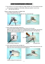
Staining
Procedure
1.
After electrophoresis, remove the gel from the cassette (page 24) and blot
proteins onto nitrocellulose membrane (page 25).
2.
Rinse the nitrocellulose membrane (containing the transferred proteins) with
deionized water for 2 minutes.
3.
Stain the nitrocellulose membrane with 20 mL of ready-to-use
InVision
™
His-tag In-gel Stain for 20 minutes at room temperature.
4.
Rinse the membrane briefly with ultrapure water.
5.
Place the membrane on a UV transilluminator equipped with a camera.
Visualize and image the membrane by exposing the membrane to UV light
form the bottom or from the top (you may place the UV transilluminator on
its side to illuminate the blot or use epi-illumination) for 4–8 seconds.
You may also use a laser-based scanner with the appropriate filters to visualize
and image the membrane.
Summary of Contents for E-PAGE Gels
Page 77: ...73...