D
IRECTION
5771498-100, R
EVISION
6
V
ENUE
™ S
ERVICE
M
ANUAL
Chapter 4 - General Procedures and Functional Checks
4-15
P R E L I M I N A R Y
4-3-3-3
Adjust the 2D Mode Controls
The following controls can be adjusted to optimize the 2D Mode display:
•
Swipe to page 2 on the Touch panel and press either
Soft
or
Sharp
Auto Tissue setting.
-
Soft
: optimizes the radial and lateral uniformity and brightness of the tissue continuously in
real-time.
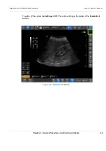
The mention “Soft” is displayed on the upper right corner of the image area
-
Sharp
: further enhances the image display by optimizing the gray scale curve.
The mention “Sharp” is displayed on the upper right corner of the image area
The Auto Tissue setting (Soft or Sharp) can be turned on/off by pressing
Auto
on the Control panel.
The last used setting is then applied.
The Auto Tissue settings are only available in live scanning and cannot be turned off when the
image is stored.
•
If available, press
Virtual Apex
(probe dependent) to improve near field imaging, allowing
increased visibility up to the width of the full probe aperture close to the surface.
•
Use the
Gain
and
TGC
controls to optimize the overall image.
Gain increases or decreases the amount of echo information displayed. TGC compensates for
depth-related attenuation in the image.
•
Use the
Depth
control to adjust the range to be imaged.
•
Use the
Modes
to toggle between the scanning system’s main modes.
•
Use the
Frequency
control (move to lower frequency) to increase penetration.
•
Press to activate the
Needle
Visualization mode. Gain, Angle and Tilt can be activated. The needle
function only applies to linear probes and the C1-5-RS convex probe.
•
Use
Split
to split the screen into two, allowing to see different, independent views on each half.
•
Use the
Reject
control to reduce noise in the image.
•
Use the
DDP
control to optimize imaging in the blood flow regions and make a cleaner, less noisy
image.
•
Use
UD Clarity
(Cardiac) or
UD Speckle reduce
(non-cardiac) to reduce image speckle. Extra care
must be taken to select the optimal Speckle reduction level, as too much filtering of speckle can
mask or obscure desired image detail.
•
Adjust
Octave
to toggle between fundamental and Harmonic mode.
•
Press
Color maps
and select a grey map from the menu on screen.
4-3-4
M Mode Checks
4-3-4-1
M-Mode Overview
This unit has three types of M-Mode:
•
Conventional M-Mode (MM): displays a distance/time plot of a cursor line in the axial plane of
the 2D-image.
•
Anatomical M-Mode (AMM): displays a distance/time plot from a cursor line, which is
independent from the axial plane. AMM is available in greyscale, color, TVI, Tissue Tracking,
Strain rate and Strain modes.
WARNING
WARNING
Always use the minimum power required to obtain acceptable
images in accordance with applicable guidelines and policies.
Summary of Contents for Venue R1
Page 477: ......


